 Movie
Movie Controller

[English] 日本語
 Yorodumi
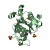
- PDB-1i04: CRYSTAL STRUCTURE OF MOUSE MAJOR URINARY PROTEIN-I FROM MOUSE LIVER -
+ Open data

- Basic information
| Entry | Database: PDB / ID: 1i04 | ||||||
|---|---|---|---|---|---|---|---|
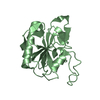
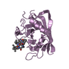
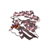
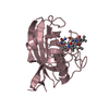
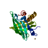
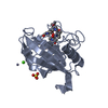
| Title | CRYSTAL STRUCTURE OF MOUSE MAJOR URINARY PROTEIN-I FROM MOUSE LIVER | ||||||
 Components Components | MAJOR URINARY PROTEIN I | ||||||
 Keywords Keywords | TRANSPORT PROTEIN / lipocalin / beta-barrel / pheromone | ||||||
| Function / homology |  Function and homology information Function and homology informationpheromone binding / negative regulation of lipid biosynthetic process / negative regulation of insulin secretion involved in cellular response to glucose stimulus / positive regulation of glucose metabolic process / energy reserve metabolic process / odorant binding / insulin receptor activity / heat generation / cellular response to lipid / positive regulation of lipid metabolic process ...pheromone binding / negative regulation of lipid biosynthetic process / negative regulation of insulin secretion involved in cellular response to glucose stimulus / positive regulation of glucose metabolic process / energy reserve metabolic process / odorant binding / insulin receptor activity / heat generation / cellular response to lipid / positive regulation of lipid metabolic process / locomotor rhythm / small molecule binding / negative regulation of lipid storage / negative regulation of gluconeogenesis / aerobic respiration / mitochondrion organization / glucose homeostasis / positive regulation of phosphatidylinositol 3-kinase/protein kinase B signal transduction / negative regulation of DNA-templated transcription / positive regulation of gene expression / : / nucleus / cytosol Similarity search - Function | ||||||
| Biological species |  | ||||||
| Method |  X-RAY DIFFRACTION / SYNCHROTRON / MOLECULAR REPLACEMENT / Resolution: 2 Å X-RAY DIFFRACTION / SYNCHROTRON / MOLECULAR REPLACEMENT / Resolution: 2 Å | ||||||
 Authors Authors | Timm, D.E. / Baker, L.J. / Mueller, H. / Zidek, L. / Novotny, M.V. | ||||||
 Citation Citation | Journal: Protein Sci. / Year: 2001 Title: Structural basis of pheromone binding to mouse major urinary protein (MUP-I) Authors: Timm, D.E. / Baker, L.J. / Mueller, H. / Zidek, L. / Novotny, M.V. | ||||||
| History |
|
- Structure visualization
| Structure viewer | Molecule: MolmilJmol/JSmol |
|---|
- Downloads & links
-Download
| PDBx/mmCIF format | 1i04.cif.gz | 47.6 KB | Display | PDBx/mmCIF format |
|---|---|---|---|---|
| PDB format | pdb1i04.ent.gz | 33.4 KB | Display | PDB format |
| PDBx/mmJSON format | 1i04.json.gz | Tree view | PDBx/mmJSON format | |
| Others |  Other downloads Other downloads |
-Validation report
| Arichive directory | https://data.pdbj.org/pub/pdb/validation_reports/i0/1i04ftp://data.pdbj.org/pub/pdb/validation_reports/i0/1i04 | HTTPS FTP |
|---|
-Related structure data
| Related structure data | 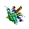 1i05C 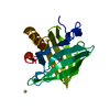 1i06C  1mupS S: Starting model for refinement C: citing same article ( |
|---|---|
| Similar structure data |


-Links
 PDBj
PDBj

- Assembly
| Deposited unit | 
| ||||||||
|---|---|---|---|---|---|---|---|---|---|
| 1 |
| ||||||||
| Unit cell |
|
-Components
| #1: Protein | Mass: 20674.396 Da / Num. of mol.: 1 / Source method: isolated from a natural source / Source: (natural) |
|---|---|
| #2: Water | ChemComp-HOH /  Mass: 18.015 Da / Num. of mol.: 154 / Source method: isolated from a natural source / Formula: H2O Mass: 18.015 Da / Num. of mol.: 154 / Source method: isolated from a natural source / Formula: H2O |
| Has protein modification | Y |
-Experimental details
-Experiment
| Experiment | Method: X-RAY DIFFRACTION / Number of used crystals: 1 |
|---|
- Sample preparation
| Crystal | Density Matthews: 2.59 Å3/Da / Density % sol: 52.54 % | |||||||||||||||
|---|---|---|---|---|---|---|---|---|---|---|---|---|---|---|---|---|
| Crystal grow | Temperature: 298 K / Method: vapor diffusion, hanging drop Details: 50-70% ammonium sulfate, citrate, phosphate buffer, VAPOR DIFFUSION, HANGING DROP, temperature 298K | |||||||||||||||
| Crystal grow | *PLUS PH range low: 7 / PH range high: 5 | |||||||||||||||
| Components of the solutions | *PLUS
|
-Data collection
| Diffraction | Mean temperature: 100 K |
|---|---|
| Diffraction source | Source: SYNCHROTRON / Site: NSLS  / Beamline: X12C / Wavelength: 1 Å / Beamline: X12C / Wavelength: 1 Å |
| Detector | Type: CUSTOM-MADE / Detector: CCD / Date: Aug 15, 1997 / Details: MIRRORS |
| Radiation | Monochromator: MIRRORS / Protocol: SINGLE WAVELENGTH / Monochromatic (M) / Laue (L): M / Scattering type: x-ray |
| Radiation wavelength | Wavelength: 1 Å / Relative weight: 1 |
| Reflection | Resolution: 2→18 Å / Num. all: 14762 / Num. obs: 14762 / % possible obs: 97.9 % / Observed criterion σ(F): 0 / Observed criterion σ(I): 0 / Redundancy: 3.3 % / Biso Wilson estimate: 25.8 Å2 / Rmerge(I) obs: 0.06 / Rsym value: 0.06 / Net I/σ(I): 17.2 |
| Reflection shell | Resolution: 2→2.07 Å / Redundancy: 3.3 % / Rmerge(I) obs: 0.276 / Mean I/σ(I) obs: 3.3 / Num. unique all: 1270 / Rsym value: 0.276 / % possible all: 85.9 |
| Reflection | *PLUS Num. measured all: 48342 / Rmerge(I) obs: 0.06 |
| Reflection shell | *PLUS % possible obs: 85.9 % |
- Processing
| Software |
| ||||||||||||||||||||
|---|---|---|---|---|---|---|---|---|---|---|---|---|---|---|---|---|---|---|---|---|---|
| Refinement | Method to determine structure: MOLECULAR REPLACEMENT Starting model: 1MUP Resolution: 2→20 Å / Cross valid method: THROUGHOUT / σ(F): 2 / σ(I): 2 / Stereochemistry target values: Engh & Huber
| ||||||||||||||||||||
| Refinement step | Cycle: LAST / Resolution: 2→20 Å
| ||||||||||||||||||||
| Refine LS restraints |
| ||||||||||||||||||||
| Software | *PLUS Name: CNS / Version: 0.9 / Classification: refinement | ||||||||||||||||||||
| Refinement | *PLUS Highest resolution: 2 Å / Lowest resolution: 20 Å / σ(F): 2 / Rfactor Rfree: 0.25 | ||||||||||||||||||||
| Solvent computation | *PLUS | ||||||||||||||||||||
| Displacement parameters | *PLUS |
