 Movie
Movie Controller

[English] 日本語
 Yorodumi
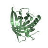
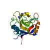
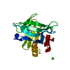
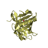
- PDB-1mup: PHEROMONE BINDING TO TWO RODENT URINARY PROTEINS REVEALED BY X-RA... -
+ Open data

- Basic information
| Entry | Database: PDB / ID: 1mup | ||||||
|---|---|---|---|---|---|---|---|
| Title | PHEROMONE BINDING TO TWO RODENT URINARY PROTEINS REVEALED BY X-RAY CRYSTALLOGRAPHY | ||||||
 Components Components | MAJOR URINARY PROTEIN | ||||||
 Keywords Keywords | PHEROMONE-BINDING | ||||||
| Function / homology |  Function and homology information Function and homology informationpheromone binding / negative regulation of lipid biosynthetic process / positive regulation of glucose metabolic process / odorant binding / energy reserve metabolic process / insulin receptor activity / negative regulation of insulin secretion involved in cellular response to glucose stimulus / heat generation / cellular response to lipid / positive regulation of lipid metabolic process ...pheromone binding / negative regulation of lipid biosynthetic process / positive regulation of glucose metabolic process / odorant binding / energy reserve metabolic process / insulin receptor activity / negative regulation of insulin secretion involved in cellular response to glucose stimulus / heat generation / cellular response to lipid / positive regulation of lipid metabolic process / locomotor rhythm / small molecule binding / negative regulation of lipid storage / negative regulation of gluconeogenesis / aerobic respiration / mitochondrion organization / glucose homeostasis / positive regulation of phosphatidylinositol 3-kinase/protein kinase B signal transduction / negative regulation of DNA-templated transcription / positive regulation of gene expression / : / nucleus / cytosol Similarity search - Function | ||||||
| Biological species |  | ||||||
| Method |  X-RAY DIFFRACTION / Resolution: 2.4 Å X-RAY DIFFRACTION / Resolution: 2.4 Å | ||||||
 Authors Authors | Bocskei, Z. / Flower, D.R. / Groom, C.R. / Phillips, S.E.V. / North, A.C.T. | ||||||
 Citation Citation | Journal: Nature / Year: 1992 Title: Pheromone binding to two rodent urinary proteins revealed by X-ray crystallography. Authors: Bocskei, Z. / Groom, C.R. / Flower, D.R. / Wright, C.E. / Phillips, S.E. / Cavaggioni, A. / Findlay, J.B. / North, A.C. #1: Journal: Experientia / Year: 1992Title: Pheromone Binding Proteins of the Mouse (Mus Musculus) Authors: Bacchini, A. / Gaetani, E. / Cavaggioni, A. #2: Journal: J.Mol.Biol. / Year: 1991Title: Crystallization of and Preliminary X-Ray Data for the Mouse Major Urinary Protein and Rat Alpha-2U Globulin Authors: Bocskei, Z. / Findlay, J.B.C. / North, A.C.T. / Phillips, S.E.V. / Somers, W.S. / Wright, C.E. / Lionetti, C. / Tirindelli, R. / Cavaggioni, A. | ||||||
| History |
|
- Structure visualization
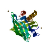
| Structure viewer | Molecule: MolmilJmol/JSmol |
|---|
- Downloads & links
-Download
| PDBx/mmCIF format | 1mup.cif.gz | 48 KB | Display | PDBx/mmCIF format |
|---|---|---|---|---|
| PDB format | pdb1mup.ent.gz | 32.4 KB | Display | PDB format |
| PDBx/mmJSON format | 1mup.json.gz | Tree view | PDBx/mmJSON format | |
| Others |  Other downloads Other downloads |
-Validation report
| Arichive directory | https://data.pdbj.org/pub/pdb/validation_reports/mu/1mupftp://data.pdbj.org/pub/pdb/validation_reports/mu/1mup | HTTPS FTP |
|---|
-Related structure data
| Similar structure data |
|---|


-Links
 PDBj
PDBj

- Assembly
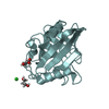
| Deposited unit | 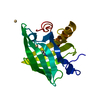
| ||||||||
|---|---|---|---|---|---|---|---|---|---|
| 1 |
| ||||||||
| Unit cell |
|
-Components
| #1: Protein | Mass: 19130.289 Da / Num. of mol.: 1 Source method: isolated from a genetically manipulated source Source: (gene. exp.) | ||||||||
|---|---|---|---|---|---|---|---|---|---|
| #2: Chemical | ChemComp-CD /   Mass: 112.411 Da / Num. of mol.: 4 / Source method: obtained synthetically / Formula: Cd Mass: 112.411 Da / Num. of mol.: 4 / Source method: obtained synthetically / Formula: Cd#3: Chemical | ChemComp-TZL / | 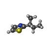  Mass: 141.234 Da / Num. of mol.: 1 / Source method: obtained synthetically / Formula: C7H11NS Mass: 141.234 Da / Num. of mol.: 1 / Source method: obtained synthetically / Formula: C7H11NS#4: Water | ChemComp-HOH / |  Mass: 18.015 Da / Num. of mol.: 77 / Source method: isolated from a natural source / Formula: H2O Mass: 18.015 Da / Num. of mol.: 77 / Source method: isolated from a natural source / Formula: H2OHas protein modification | Y | Nonpolymer details | THE ENTRY INCLUDES FOUR CADMIUM IONS WITH VARYING OCCUPANCIE | |
-Experimental details
-Experiment
| Experiment | Method: X-RAY DIFFRACTION |
|---|
- Sample preparation
| Crystal | Density Matthews: 2.36 Å3/Da / Density % sol: 47.87 % | ||||||||||||||||||||||||||||||||||||||||||
|---|---|---|---|---|---|---|---|---|---|---|---|---|---|---|---|---|---|---|---|---|---|---|---|---|---|---|---|---|---|---|---|---|---|---|---|---|---|---|---|---|---|---|---|
| Crystal grow | *PLUS pH: 5.5 / Method: vapor diffusion, hanging dropDetails: taken from Bocskei, Z. et al (1991). J. Mol. Biol., 218, 699-701. | ||||||||||||||||||||||||||||||||||||||||||
| Components of the solutions | *PLUS
|
-Data collection
| Radiation | Scattering type: x-ray |
|---|---|
| Radiation wavelength | Relative weight: 1 |
- Processing
| Software |
| ||||||||||||||||||||||||||||||||||||||||||||||||||||||||||||||||||||||||||||||||
|---|---|---|---|---|---|---|---|---|---|---|---|---|---|---|---|---|---|---|---|---|---|---|---|---|---|---|---|---|---|---|---|---|---|---|---|---|---|---|---|---|---|---|---|---|---|---|---|---|---|---|---|---|---|---|---|---|---|---|---|---|---|---|---|---|---|---|---|---|---|---|---|---|---|---|---|---|---|---|---|---|---|
| Refinement | Resolution: 2.4→14 Å / Rfactor Rwork: 0.191 / Rfactor obs: 0.191 | ||||||||||||||||||||||||||||||||||||||||||||||||||||||||||||||||||||||||||||||||
| Refinement step | Cycle: LAST / Resolution: 2.4→14 Å
| ||||||||||||||||||||||||||||||||||||||||||||||||||||||||||||||||||||||||||||||||
| Refine LS restraints |
| ||||||||||||||||||||||||||||||||||||||||||||||||||||||||||||||||||||||||||||||||
| Software | *PLUS Name: TNT / Classification: refinement | ||||||||||||||||||||||||||||||||||||||||||||||||||||||||||||||||||||||||||||||||
| Refinement | *PLUS Highest resolution: 2.4 Å / Lowest resolution: 14 Å / Rfactor obs: 0.191 | ||||||||||||||||||||||||||||||||||||||||||||||||||||||||||||||||||||||||||||||||
| Solvent computation | *PLUS | ||||||||||||||||||||||||||||||||||||||||||||||||||||||||||||||||||||||||||||||||
| Displacement parameters | *PLUS | ||||||||||||||||||||||||||||||||||||||||||||||||||||||||||||||||||||||||||||||||
| Refine LS restraints | *PLUS
|
