 Movie
Movie Controller

[English] 日本語
 Yorodumi
- PDB-1hyo: CRYSTAL STRUCTURE OF FUMARYLACETOACETATE HYDROLASE COMPLEXED WITH... -
+ Open data

- Basic information
| Entry | Database: PDB / ID: 1hyo | ||||||
|---|---|---|---|---|---|---|---|
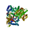
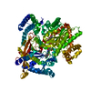
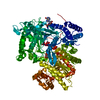
| Title | CRYSTAL STRUCTURE OF FUMARYLACETOACETATE HYDROLASE COMPLEXED WITH 4-(HYDROXYMETHYLPHOSPHINOYL)-3-OXO-BUTANOIC ACID | ||||||
 Components Components | FUMARYLACETOACETATE HYDROLASE | ||||||
 Keywords Keywords | HYDROLASE / beta-sandwich roll | ||||||
| Function / homology |  Function and homology information Function and homology informationTyrosine catabolism / fumarylacetoacetase / fumarylacetoacetase activity / homogentisate catabolic process / L-tyrosine catabolic process / L-phenylalanine catabolic process / L-arginine catabolic process / lipid metabolic process / metal ion binding Similarity search - Function | ||||||
| Biological species |  | ||||||
| Method |  X-RAY DIFFRACTION / SYNCHROTRON / MOLECULAR REPLACEMENT / Resolution: 1.3 Å X-RAY DIFFRACTION / SYNCHROTRON / MOLECULAR REPLACEMENT / Resolution: 1.3 Å | ||||||
 Authors Authors | Bateman, R.L. / Bhanumoorthy, P. / Witte, J.F. / McClard, R.W. / Grompe, M. / Timm, D.E. | ||||||
 Citation Citation | Journal: J.Biol.Chem. / Year: 2001 Title: Mechanistic inferences from the crystal structure of fumarylacetoacetate hydrolase with a bound phosphorus-based inhibitor. Authors: Bateman, R.L. / Bhanumoorthy, P. / Witte, J.F. / McClard, R.W. / Grompe, M. / Timm, D.E. | ||||||
| History |
|
- Structure visualization
| Structure viewer | Molecule: MolmilJmol/JSmol |
|---|
- Downloads & links
-Download
| PDBx/mmCIF format | 1hyo.cif.gz | 195.6 KB | Display | PDBx/mmCIF format |
|---|---|---|---|---|
| PDB format | pdb1hyo.ent.gz | 150.5 KB | Display | PDB format |
| PDBx/mmJSON format | 1hyo.json.gz | Tree view | PDBx/mmJSON format | |
| Others |  Other downloads Other downloads |
-Validation report
| Arichive directory | https://data.pdbj.org/pub/pdb/validation_reports/hy/1hyoftp://data.pdbj.org/pub/pdb/validation_reports/hy/1hyo | HTTPS FTP |
|---|
-Related structure data
| Related structure data |  1qcnS S: Starting model for refinement |
|---|---|
| Similar structure data |


-Links
 PDBj
PDBj- Assembly
| Deposited unit | 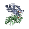
| ||||||||
|---|---|---|---|---|---|---|---|---|---|
| 1 |
| ||||||||
| Unit cell |
|
-Components
-Protein , 1 types, 2 molecules AB
| #1: Protein | Mass: 46296.660 Da / Num. of mol.: 2 Source method: isolated from a genetically manipulated source Source: (gene. exp.)  |
|---|
-Non-polymers , 6 types, 843 molecules 


| #2: Chemical |  Mass: 24.305 Da / Num. of mol.: 2 / Source method: obtained synthetically / Formula: Mg Mass: 24.305 Da / Num. of mol.: 2 / Source method: obtained synthetically / Formula: Mg#3: Chemical |  Mass: 40.078 Da / Num. of mol.: 2 / Source method: obtained synthetically / Formula: Ca Mass: 40.078 Da / Num. of mol.: 2 / Source method: obtained synthetically / Formula: Ca#4: Chemical |  Mass: 59.044 Da / Num. of mol.: 2 / Source method: obtained synthetically / Formula: C2H3O2 Mass: 59.044 Da / Num. of mol.: 2 / Source method: obtained synthetically / Formula: C2H3O2#5: Chemical |  Mass: 180.096 Da / Num. of mol.: 2 / Source method: obtained synthetically / Formula: C5H9O5P Mass: 180.096 Da / Num. of mol.: 2 / Source method: obtained synthetically / Formula: C5H9O5P#6: Chemical | ChemComp-NI / |  Mass: 58.693 Da / Num. of mol.: 1 / Source method: obtained synthetically / Formula: Ni Mass: 58.693 Da / Num. of mol.: 1 / Source method: obtained synthetically / Formula: Ni#7: Water | ChemComp-HOH / | Mass: 18.015 Da / Num. of mol.: 834 / Source method: isolated from a natural source / Formula: H2O |
|---|
-Experimental details
-Experiment
| Experiment | Method: X-RAY DIFFRACTION / Number of used crystals: 1 |
|---|
- Sample preparation
| Crystal | Density Matthews: 2.49 Å3/Da / Density % sol: 50.7 % | ||||||||||||||||||||||||||||||
|---|---|---|---|---|---|---|---|---|---|---|---|---|---|---|---|---|---|---|---|---|---|---|---|---|---|---|---|---|---|---|---|
| Crystal grow | Temperature: 298 K / Method: vapor diffusion, hanging drop / pH: 6.5 Details: PEG 6000, nickel acetate, cacodylate, pH 6.5, VAPOR DIFFUSION, HANGING DROP, temperature 298K | ||||||||||||||||||||||||||||||
| Crystal grow | *PLUS | ||||||||||||||||||||||||||||||
| Components of the solutions | *PLUS
|
-Data collection
| Diffraction | Mean temperature: 100 K |
|---|---|
| Diffraction source | Source: SYNCHROTRON / Site: NSLS  / Beamline: X12C / Wavelength: 0.98 Å / Beamline: X12C / Wavelength: 0.98 Å |
| Detector | Type: BRANDEIS - B4 / Detector: CCD / Date: Nov 15, 1998 / Details: mirrors |
| Radiation | Monochromator: mirrors / Protocol: SINGLE WAVELENGTH / Monochromatic (M) / Laue (L): M / Scattering type: x-ray |
| Radiation wavelength | Wavelength: 0.98 Å / Relative weight: 1 |
| Reflection | Resolution: 1.3→27 Å / Num. all: 235526 / Num. obs: 227518 / % possible obs: 96.6 % / Observed criterion σ(F): 2 / Observed criterion σ(I): 2 / Redundancy: 2.95 % / Biso Wilson estimate: 13.4 Å2 / Rmerge(I) obs: 0.046 / Rsym value: 0.046 / Net I/σ(I): 15.6 |
| Reflection shell | Resolution: 1.27→1.32 Å / Redundancy: 2.95 % / Rmerge(I) obs: 0.366 / Mean I/σ(I) obs: 1.4 / Num. unique all: 19224 / Rsym value: 0.366 / % possible all: 81.8 |
| Reflection | *PLUS Num. measured all: 672278 |
| Reflection shell | *PLUS % possible obs: 81.8 % |
- Processing
| Software |
| |||||||||||||||||||||||||
|---|---|---|---|---|---|---|---|---|---|---|---|---|---|---|---|---|---|---|---|---|---|---|---|---|---|---|
| Refinement | Method to determine structure: MOLECULAR REPLACEMENT Starting model: PDB ENTRY 1QCN Resolution: 1.3→27 Å / Isotropic thermal model: isotropic / Cross valid method: THROUGHOUT / σ(F): 0 / σ(I): 0 / Stereochemistry target values: Protin
| |||||||||||||||||||||||||
| Displacement parameters | Biso mean: 13.4 Å2 | |||||||||||||||||||||||||
| Refine analyze | Luzzati coordinate error obs: 0.047 Å | |||||||||||||||||||||||||
| Refinement step | Cycle: LAST / Resolution: 1.3→27 Å
| |||||||||||||||||||||||||
| Refine LS restraints |
| |||||||||||||||||||||||||
| LS refinement shell | Resolution: 1.3→1.36 Å /
| |||||||||||||||||||||||||
| Software | *PLUS Name: REFMAC / Classification: refinement | |||||||||||||||||||||||||
| Refinement | *PLUS Highest resolution: 1.3 Å / σ(F): 0 | |||||||||||||||||||||||||
| Solvent computation | *PLUS | |||||||||||||||||||||||||
| Displacement parameters | *PLUS Biso mean: 13.4 Å2 | |||||||||||||||||||||||||
| Refine LS restraints | *PLUS
| |||||||||||||||||||||||||
| LS refinement shell | *PLUS Rfactor obs: 0.234 |
