 Movie
Movie Controller

[English] 日本語
 Yorodumi
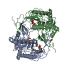
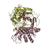
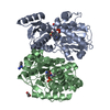
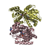
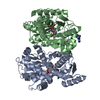
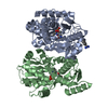
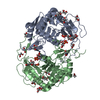
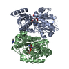
- PDB-1hxq: THE STRUCTURE OF NUCLEOTIDYLATED GALACTOSE-1-PHOSPHATE URIDYLYLTR... -
+ Open data

- Basic information
| Entry | Database: PDB / ID: 1hxq | ||||||
|---|---|---|---|---|---|---|---|
| Title | THE STRUCTURE OF NUCLEOTIDYLATED GALACTOSE-1-PHOSPHATE URIDYLYLTRANSFERASE FROM ESCHERICHIA COLI AT 1.86 ANGSTROMS RESOLUTION | ||||||
 Components Components | HEXOSE-1-PHOSPHATE URIDYLYLTRANSFERASE | ||||||
 Keywords Keywords | NUCLEOTIDYLTRANSFERASE / METALLOENZYME / NUCLEOTIDYLATED HISTIDINE / REACTION INTERMEDIATE | ||||||
| Function / homology |  Function and homology information Function and homology informationUDP-glucose-hexose-1-phosphate uridylyltransferase / UDP-glucose:hexose-1-phosphate uridylyltransferase activity / galactokinase activity / beta-D-galactose catabolic process via UDP-galactose, Leloir pathway / ferrous iron binding / zinc ion binding / cytosol / cytoplasm Similarity search - Function | ||||||
| Biological species |  | ||||||
| Method |  X-RAY DIFFRACTION / Resolution: 1.86 Å X-RAY DIFFRACTION / Resolution: 1.86 Å | ||||||
 Authors Authors | Wedekind, J.E. / Frey, P.A. / Rayment, I. | ||||||
 Citation Citation | Journal: Biochemistry / Year: 1996 Title: The structure of nucleotidylated histidine-166 of galactose-1-phosphate uridylyltransferase provides insight into phosphoryl group transfer. Authors: Wedekind, J.E. / Frey, P.A. / Rayment, I. #1: Journal: Biochemistry / Year: 1995Title: Three-Dimensional Structure of Galactose-1-Phosphate Uridylyltransferase from Escherichia Coli at 1.8 A Resolution Authors: Wedekind, J.E. / Frey, P.A. / Rayment, I. #2: Journal: Biochemistry / Year: 1995Title: Galactose-1-Phosphate Uridylyltransferase from Escherichia Coli, a Zinc and Iron Metalloenzyme Authors: Ruzicka, F.J. / Wedekind, J.E. / Kim, J. / Rayment, I. / Frey, P.A. #3: Journal: Acta Crystallogr.,Sect.D / Year: 1994Title: Crystallization and Preliminary Crystallographic Analysis of Galactose-1-Phosphate Uridylyltransferase from Escherichia Coli Authors: Wedekind, J.E. / Frey, P.A. / Rayment, I. #4: Journal: Nucleic Acids Res. / Year: 1987Title: The Nucleotide Sequence of the Gal T Gene of Escherichia Coli Authors: Cornwell, T.L. / Adhya, S.L. / Reznikoff, W.S. / Frey, P.A. | ||||||
| History |
|
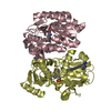
- Structure visualization
| Structure viewer | Molecule: MolmilJmol/JSmol |
|---|
- Downloads & links
-Download
| PDBx/mmCIF format | 1hxq.cif.gz | 166.1 KB | Display | PDBx/mmCIF format |
|---|---|---|---|---|
| PDB format | pdb1hxq.ent.gz | 129.8 KB | Display | PDB format |
| PDBx/mmJSON format | 1hxq.json.gz | Tree view | PDBx/mmJSON format | |
| Others |  Other downloads Other downloads |
-Validation report
| Arichive directory | https://data.pdbj.org/pub/pdb/validation_reports/hx/1hxqftp://data.pdbj.org/pub/pdb/validation_reports/hx/1hxq | HTTPS FTP |
|---|
-Related structure data
| Similar structure data |
|---|
-Links
 PDBj
PDBj


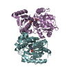
- Assembly
| Deposited unit | 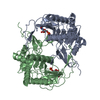
| ||||||||
|---|---|---|---|---|---|---|---|---|---|
| 1 |
| ||||||||
| Unit cell |
| ||||||||
| Noncrystallographic symmetry (NCS) | NCS oper: (Code: given Matrix: (0.94373, -0.00281, 0.33071), Vector: Details | THE CRYSTALLOGRAPHICALLY INDEPENDENT UNIT IS ONE DIMER OF CHEMICALLY IDENTICAL SUBUNITS. | |
-Components
| #1: Protein | Mass: 39694.598 Da / Num. of mol.: 2 Source method: isolated from a genetically manipulated source Source: (gene. exp.) References: UniProt: P09148, UDP-glucose-hexose-1-phosphate uridylyltransferase #2: Chemical |   Mass: 65.409 Da / Num. of mol.: 2 / Source method: obtained synthetically / Formula: Zn Mass: 65.409 Da / Num. of mol.: 2 / Source method: obtained synthetically / Formula: Zn#3: Chemical |   Mass: 55.845 Da / Num. of mol.: 2 / Source method: obtained synthetically / Formula: Fe Mass: 55.845 Da / Num. of mol.: 2 / Source method: obtained synthetically / Formula: Fe#4: Chemical |   Mass: 324.181 Da / Num. of mol.: 2 / Source method: obtained synthetically / Formula: C9H13N2O9P Mass: 324.181 Da / Num. of mol.: 2 / Source method: obtained synthetically / Formula: C9H13N2O9P#5: Water | ChemComp-HOH / |  Mass: 18.015 Da / Num. of mol.: 730 / Source method: isolated from a natural source / Formula: H2O Mass: 18.015 Da / Num. of mol.: 730 / Source method: isolated from a natural source / Formula: H2OCompound details | GLN 168 MAKES CLOSE CONTACTS TO O2 OF THE U5P PHOSPHORYL GROUP. MUTATION OF THIS RESIDUE TO ARG HAS ...GLN 168 MAKES CLOSE CONTACTS TO O2 OF THE U5P PHOSPHORYL | Has protein modification | Y | Nonpolymer details | THIS ENTRY REPRESENTS THE STRUCTURE OF GALACTOSE-1-PHOSPHATE URIDYLYLTRANSFERASE IN WHICH NE2 OF ...THIS ENTRY REPRESENTS | |
|---|
-Experimental details
-Experiment
| Experiment | Method: X-RAY DIFFRACTION / Number of used crystals: 2 |
|---|
- Sample preparation
| Crystal | Density Matthews: 2.77 Å3/Da / Density % sol: 55.6 % | ||||||||||||||||||||||||||||||
|---|---|---|---|---|---|---|---|---|---|---|---|---|---|---|---|---|---|---|---|---|---|---|---|---|---|---|---|---|---|---|---|
| Crystal grow | pH: 7.1 Details: EACH CRYSTAL WAS REACTED WITH SUBSTRATE UDP-GLUCOSE IN A SYNTHETIC MOTHER LIQUOR DEVOID OF LITHIUM SULFATE AT PH 7.1. | ||||||||||||||||||||||||||||||
| Crystal grow | *PLUS pH: 6 / Method: vapor diffusion | ||||||||||||||||||||||||||||||
| Components of the solutions | *PLUS
|
-Data collection
| Diffraction | Mean temperature: 113 K |
|---|---|
| Diffraction source | Wavelength: 1.5418 |
| Detector | Type: SIEMENS HI-STAR / Detector: AREA DETECTOR / Date: 1995 / Details: SUPPER DOUBLE-FOCUSING MIRRORS WITH NICKEL FOIL |
| Radiation | Monochromatic (M) / Laue (L): M / Scattering type: x-ray |
| Radiation wavelength | Wavelength: 1.5418 Å / Relative weight: 1 |
| Reflection | Resolution: 1.86→65 Å / Num. obs: 70581 / % possible obs: 97 % / Observed criterion σ(I): -1 / Redundancy: 2.5 % / Biso Wilson estimate: 19.1 Å2 / Rmerge(I) obs: 0.063 |
| Reflection | *PLUS Num. all: 15045 / Num. measured all: 206202 / Rmerge(I) obs: 0.0625 |
| Reflection shell | *PLUS Highest resolution: 1.86 Å / % possible obs: 90 % / Num. unique obs: 10989 / Num. measured obs: 19085 / Rmerge(I) obs: 0.165 |
- Processing
| Software |
| ||||||||||||||||||||||||||||||||||||||||||||||||||
|---|---|---|---|---|---|---|---|---|---|---|---|---|---|---|---|---|---|---|---|---|---|---|---|---|---|---|---|---|---|---|---|---|---|---|---|---|---|---|---|---|---|---|---|---|---|---|---|---|---|---|---|
| Refinement | Resolution: 1.86→65 Å / σ(F): 0 Details: REGIONS A 36 THROUGH A 44 AND B 31 THROUGH B 43 ARE DISORDERED. NO SIGNIFICANT ELECTRON DENSITY CAN BE SEEN FOR THESE REGIONS AT THE RESOLUTION OF THE REFINED MODEL. RESIDUES B 347 AND B 348 ...Details: REGIONS A 36 THROUGH A 44 AND B 31 THROUGH B 43 ARE DISORDERED. NO SIGNIFICANT ELECTRON DENSITY CAN BE SEEN FOR THESE REGIONS AT THE RESOLUTION OF THE REFINED MODEL. RESIDUES B 347 AND B 348 OF SUBUNIT II ARE DISORDERED AS WELL.
| ||||||||||||||||||||||||||||||||||||||||||||||||||
| Solvent computation | Solvent model: TNT / Bsol: 526 Å2 / ksol: 0.7 e/Å3 | ||||||||||||||||||||||||||||||||||||||||||||||||||
| Refinement step | Cycle: LAST / Resolution: 1.86→65 Å
| ||||||||||||||||||||||||||||||||||||||||||||||||||
| Refine LS restraints |
| ||||||||||||||||||||||||||||||||||||||||||||||||||
| Software | *PLUS Name: TNT / Version: 5-E / Classification: refinement | ||||||||||||||||||||||||||||||||||||||||||||||||||
| Refinement | *PLUS Rfactor all: 0.196 | ||||||||||||||||||||||||||||||||||||||||||||||||||
| Solvent computation | *PLUS | ||||||||||||||||||||||||||||||||||||||||||||||||||
| Displacement parameters | *PLUS | ||||||||||||||||||||||||||||||||||||||||||||||||||
| Refine LS restraints | *PLUS
|
