 Movie
Movie Controller


+ Open data

- Basic information
| Entry | Database: PDB / ID: 1gup | ||||||
|---|---|---|---|---|---|---|---|
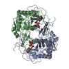
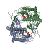
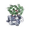
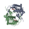
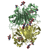
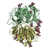
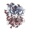
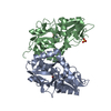
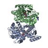
| Title | STRUCTURE OF NUCLEOTIDYLTRANSFERASE COMPLEXED WITH UDP-GALACTOSE | ||||||
 Components Components | GALACTOSE-1-PHOSPHATE URIDYLYLTRANSFERASE | ||||||
 Keywords Keywords | NUCLEOTIDYLTRANSFERASE / TRANSFERASE / GALACTOSE METABOLISM | ||||||
| Function / homology |  Function and homology information Function and homology informationUDP-glucose-hexose-1-phosphate uridylyltransferase / UDP-glucose:hexose-1-phosphate uridylyltransferase activity / galactokinase activity / beta-D-galactose catabolic process via UDP-galactose, Leloir pathway / ferrous iron binding / zinc ion binding / cytoplasm / cytosol Similarity search - Function | ||||||
| Biological species |  | ||||||
| Method |  X-RAY DIFFRACTION / Resolution: 1.8 Å X-RAY DIFFRACTION / Resolution: 1.8 Å | ||||||
 Authors Authors | Thoden, J.B. / Rayment, I. / Holden, H. | ||||||
 Citation Citation | Journal: Biochemistry / Year: 1997 Title: Structural analysis of the H166G site-directed mutant of galactose-1-phosphate uridylyltransferase complexed with either UDP-glucose or UDP-galactose: detailed description of the nucleotide sugar binding site. Authors: Thoden, J.B. / Ruzicka, F.J. / Frey, P.A. / Rayment, I. / Holden, H.M. | ||||||
| History |
|
- Structure visualization
| Structure viewer | Molecule: MolmilJmol/JSmol |
|---|
- Downloads & links
-Download
| PDBx/mmCIF format | 1gup.cif.gz | 321.9 KB | Display | PDBx/mmCIF format |
|---|---|---|---|---|
| PDB format | pdb1gup.ent.gz | 256.5 KB | Display | PDB format |
| PDBx/mmJSON format | 1gup.json.gz | Tree view | PDBx/mmJSON format | |
| Others |  Other downloads Other downloads |
-Validation report
| Arichive directory | https://data.pdbj.org/pub/pdb/validation_reports/gu/1gupftp://data.pdbj.org/pub/pdb/validation_reports/gu/1gup | HTTPS FTP |
|---|
-Related structure data


-Links
 PDBj
PDBj


- Assembly
| Deposited unit | 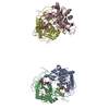
| ||||||||
|---|---|---|---|---|---|---|---|---|---|
| 1 | 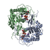
| ||||||||
| 2 | 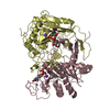
| ||||||||
| Unit cell |
|
-Components
-Protein , 1 types, 4 molecules ABCD
| #1: Protein | Mass: 39613.500 Da / Num. of mol.: 4 / Mutation: H166G Source method: isolated from a genetically manipulated source Source: (gene. exp.) |
|---|
-Non-polymers , 5 types, 1265 molecules 


| #2: Chemical | ChemComp-GDU /  Mass: 566.302 Da / Num. of mol.: 4 Mass: 566.302 Da / Num. of mol.: 4Source method: isolated from a genetically manipulated source Formula: C15H24N2O17P2 #3: Chemical | ChemComp-ZN /  Mass: 65.409 Da / Num. of mol.: 4 / Source method: obtained synthetically / Formula: Zn Mass: 65.409 Da / Num. of mol.: 4 / Source method: obtained synthetically / Formula: Zn#4: Chemical | ChemComp-FE /  Mass: 55.845 Da / Num. of mol.: 4 / Source method: obtained synthetically / Formula: Fe Mass: 55.845 Da / Num. of mol.: 4 / Source method: obtained synthetically / Formula: Fe#5: Chemical | ChemComp-K /  Mass: 39.098 Da / Num. of mol.: 4 / Source method: obtained synthetically / Formula: K Mass: 39.098 Da / Num. of mol.: 4 / Source method: obtained synthetically / Formula: K#6: Water | ChemComp-HOH / | Mass: 18.015 Da / Num. of mol.: 1249 / Source method: isolated from a natural source / Formula: H2O |
|---|
-Experimental details
-Experiment
| Experiment | Method: X-RAY DIFFRACTION |
|---|
- Sample preparation
| Crystal | Density Matthews: 2.32 Å3/Da / Density % sol: 47.04 % | ||||||||||||||||||||||||||||||||||||||||||
|---|---|---|---|---|---|---|---|---|---|---|---|---|---|---|---|---|---|---|---|---|---|---|---|---|---|---|---|---|---|---|---|---|---|---|---|---|---|---|---|---|---|---|---|
| Crystal grow | *PLUS Temperature: 4 ℃ / pH: 8 / Method: vapor diffusion, hanging drop | ||||||||||||||||||||||||||||||||||||||||||
| Components of the solutions | *PLUS
|
-Data collection
| Diffraction | Mean temperature: 123 K |
|---|---|
| Detector | Type: BRUKER NONIUS HI-STAR / Detector: MULTIWIRE AREA DETECTOR / Details: SUPER LONG DOUBLE-FOCUSING MIRRORS |
| Radiation | Scattering type: x-ray |
| Radiation wavelength | Relative weight: 1 |
| Reflection | Num. obs: 123193 / % possible obs: 90 % |
| Reflection | *PLUS Highest resolution: 1.8 Å / Lowest resolution: 30 Å / Num. measured all: 244754 / Rmerge(I) obs: 0.047 |
| Reflection shell | *PLUS Highest resolution: 1.8 Å / % possible obs: 83 % / Num. unique obs: 13689 / Num. measured obs: 20254 / Rmerge(I) obs: 0.099 |
- Processing
| Software |
| ||||||||||||||||||||||||||||||
|---|---|---|---|---|---|---|---|---|---|---|---|---|---|---|---|---|---|---|---|---|---|---|---|---|---|---|---|---|---|---|---|
| Refinement | Resolution: 1.8→30 Å /
| ||||||||||||||||||||||||||||||
| Refinement step | Cycle: LAST / Resolution: 1.8→30 Å
| ||||||||||||||||||||||||||||||
| Refine LS restraints |
| ||||||||||||||||||||||||||||||
| Software | *PLUS Name: TNT / Classification: refinement | ||||||||||||||||||||||||||||||
| Refinement | *PLUS Rfactor obs: 0.191 | ||||||||||||||||||||||||||||||
| Solvent computation | *PLUS | ||||||||||||||||||||||||||||||
| Displacement parameters | *PLUS | ||||||||||||||||||||||||||||||
| Refine LS restraints | *PLUS
|
