 Movie
Movie Controller

[English] 日本語
 Yorodumi
- PDB-5m4z: Crystal structure of the complex of T.spiralis thymidylate syntha... -
+ Open data

- Basic information
| Entry | Database: PDB / ID: 5m4z | ||||||
|---|---|---|---|---|---|---|---|
| Title | Crystal structure of the complex of T.spiralis thymidylate synthase with N(4)-hydroxy-2'-deoxycytidine-5'-monophosphate, crystallized in the presence of N(5,10)-methylenetetrahydrofolate | ||||||
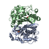
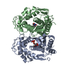
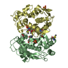
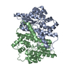
 Components Components | Thymidylate synthase | ||||||
 Keywords Keywords | TRANSFERASE / enzyme-inhibitor complex / high-resolution structure / anticancer target | ||||||
| Function / homology |  Function and homology information Function and homology informationthymidylate synthase / thymidylate synthase activity / dTTP biosynthetic process / dTMP biosynthetic process / methylation / mitochondrion / cytosol Similarity search - Function | ||||||
| Biological species |  Trichinella spiralis (invertebrata) Trichinella spiralis (invertebrata) | ||||||
| Method |  X-RAY DIFFRACTION / SYNCHROTRON / MOLECULAR REPLACEMENT / Resolution: 1.179 Å X-RAY DIFFRACTION / SYNCHROTRON / MOLECULAR REPLACEMENT / Resolution: 1.179 Å | ||||||
 Authors Authors | Wilk, P. / Maj, P. / Jarmula, A. / Dowiercial, A. / Rode, W. | ||||||
 Citation Citation | Journal: Int J Mol Sci / Year: 2021 Title: Molecular Mechanism of Thymidylate Synthase Inhibition by N 4 -Hydroxy-dCMP in View of Spectrophotometric and Crystallographic Studies. Authors: Maj, P. / Jarmula, A. / Wilk, P. / Prokopowicz, M. / Rypniewski, W. / Zielinski, Z. / Dowiercial, A. / Bzowska, A. / Rode, W. | ||||||
| History |
|
- Structure visualization
| Structure viewer | Molecule: MolmilJmol/JSmol |
|---|
- Downloads & links
-Download
| PDBx/mmCIF format | 5m4z.cif.gz | 359 KB | Display | PDBx/mmCIF format |
|---|---|---|---|---|
| PDB format | pdb5m4z.ent.gz | 299 KB | Display | PDB format |
| PDBx/mmJSON format | 5m4z.json.gz | Tree view | PDBx/mmJSON format | |
| Others |  Other downloads Other downloads |
-Validation report
| Arichive directory | https://data.pdbj.org/pub/pdb/validation_reports/m4/5m4zftp://data.pdbj.org/pub/pdb/validation_reports/m4/5m4z | HTTPS FTP |
|---|
-Related structure data
| Related structure data |  6f6zC  4g9u S: Starting model for refinement C: citing same article ( |
|---|---|
| Similar structure data |

-Links
 PDBj
PDBj- Assembly
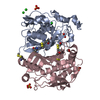
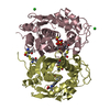
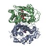
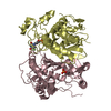
| Deposited unit | 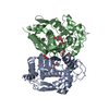
| ||||||||
|---|---|---|---|---|---|---|---|---|---|
| 1 |
| ||||||||
| Unit cell |
|
-Components
| #1: Protein | Mass: 37240.371 Da / Num. of mol.: 2 Source method: isolated from a genetically manipulated source Source: (gene. exp.) Trichinella spiralis (invertebrata) / Gene: ts, Tsp_03568 / Production host:  #2: Chemical | 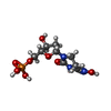  Mass: 325.212 Da / Num. of mol.: 2 / Source method: obtained synthetically / Formula: C9H16N3O8P Mass: 325.212 Da / Num. of mol.: 2 / Source method: obtained synthetically / Formula: C9H16N3O8P#3: Chemical | ChemComp-GOL /   Mass: 92.094 Da / Num. of mol.: 5 / Source method: obtained synthetically / Formula: C3H8O3 Mass: 92.094 Da / Num. of mol.: 5 / Source method: obtained synthetically / Formula: C3H8O3#4: Water | ChemComp-HOH / |  Mass: 18.015 Da / Num. of mol.: 291 / Source method: isolated from a natural source / Formula: H2O Mass: 18.015 Da / Num. of mol.: 291 / Source method: isolated from a natural source / Formula: H2OHas protein modification | Y | |
|---|
-Experimental details
-Experiment
| Experiment | Method: X-RAY DIFFRACTION / Number of used crystals: 1 |
|---|
- Sample preparation
| Crystal | Density Matthews: 2.07 Å3/Da / Density % sol: 40.46 % |
|---|---|
| Crystal grow | Temperature: 277 K / Method: vapor diffusion, hanging drop / pH: 7.1 / Details: 0.1M NaF pH 7.1, 19% PEG 3350, 0.02% NaN3 |
-Data collection
| Diffraction | Mean temperature: 100 K |
|---|---|
| Diffraction source | Source: SYNCHROTRON / Site: BESSY  / Beamline: 14.1 / Wavelength: 0.918 Å / Beamline: 14.1 / Wavelength: 0.918 Å |
| Detector | Type: DECTRIS PILATUS 6M / Detector: PIXEL / Date: May 15, 2015 |
| Radiation | Protocol: SINGLE WAVELENGTH / Monochromatic (M) / Laue (L): M / Scattering type: x-ray |
| Radiation wavelength | Wavelength: 0.918 Å / Relative weight: 1 |
| Reflection | Resolution: 1.179→28.092 Å / Num. obs: 172936 / % possible obs: 97.41 % / Redundancy: 3.61 % / CC1/2: 1 / Rmerge(I) obs: 0.034 / Rsym value: 0.04 / Net I/σ(I): 16.43 |
| Reflection shell | Resolution: 1.179→1.21 Å / % possible all: 89 |
- Processing
| Software |
| ||||||||||||||||||||||||||||||||||||||||||||||||||||||||||||||||||||||||||||||||||||||||||||||||||
|---|---|---|---|---|---|---|---|---|---|---|---|---|---|---|---|---|---|---|---|---|---|---|---|---|---|---|---|---|---|---|---|---|---|---|---|---|---|---|---|---|---|---|---|---|---|---|---|---|---|---|---|---|---|---|---|---|---|---|---|---|---|---|---|---|---|---|---|---|---|---|---|---|---|---|---|---|---|---|---|---|---|---|---|---|---|---|---|---|---|---|---|---|---|---|---|---|---|---|---|
| Refinement | Method to determine structure: MOLECULAR REPLACEMENT Starting model: 4g9u 4g9u Resolution: 1.179→28.092 Å / SU ML: 0.1 / Cross valid method: FREE R-VALUE / σ(F): 1.97 / Phase error: 17.22
| ||||||||||||||||||||||||||||||||||||||||||||||||||||||||||||||||||||||||||||||||||||||||||||||||||
| Solvent computation | Shrinkage radii: 0.9 Å / VDW probe radii: 1.11 Å | ||||||||||||||||||||||||||||||||||||||||||||||||||||||||||||||||||||||||||||||||||||||||||||||||||
| Displacement parameters | Biso max: 130.28 Å2 / Biso mean: 23.8067 Å2 / Biso min: 8.17 Å2 | ||||||||||||||||||||||||||||||||||||||||||||||||||||||||||||||||||||||||||||||||||||||||||||||||||
| Refinement step | Cycle: final / Resolution: 1.179→28.092 Å
| ||||||||||||||||||||||||||||||||||||||||||||||||||||||||||||||||||||||||||||||||||||||||||||||||||
| Refine LS restraints |
| ||||||||||||||||||||||||||||||||||||||||||||||||||||||||||||||||||||||||||||||||||||||||||||||||||
| LS refinement shell | Refine-ID: X-RAY DIFFRACTION / Total num. of bins used: 13
|
