 Movie
Movie Controller


+ Open data

- Basic information
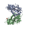
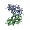
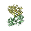
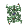
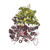
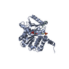
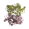
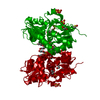
| Entry | Database: PDB / ID: 4yki | ||||||
|---|---|---|---|---|---|---|---|
| Title | Mnemiopsis leidyi ML032222a iGluR LBD glycine complex | ||||||
 Components Components | ML032222a iGluR | ||||||
 Keywords Keywords | MEMBRANE PROTEIN / Glutamate Receptor / Ion Channel | ||||||
| Function / homology |  Function and homology information Function and homology informationorganelle / ligand-gated monoatomic ion channel activity / metal ion binding / plasma membrane Similarity search - Function | ||||||
| Biological species |  Mnemiopsis leidyi (sea walnut) Mnemiopsis leidyi (sea walnut) | ||||||
| Method |  X-RAY DIFFRACTION / SYNCHROTRON / MOLECULAR REPLACEMENT / Resolution: 1.21 Å X-RAY DIFFRACTION / SYNCHROTRON / MOLECULAR REPLACEMENT / Resolution: 1.21 Å | ||||||
 Authors Authors | Alberstein, R.G. / Mayer, M.L. | ||||||
 Citation Citation | Journal: Proc.Natl.Acad.Sci.USA / Year: 2015 Title: Glycine activated ion channel subunits encoded by ctenophore glutamate receptor genes. Authors: Alberstein, R. / Grey, R. / Zimmet, A. / Simmons, D.K. / Mayer, M.L. | ||||||
| History |
|
- Structure visualization
| Structure viewer | Molecule: MolmilJmol/JSmol |
|---|
- Downloads & links
-Download
| PDBx/mmCIF format | 4yki.cif.gz | 293.7 KB | Display | PDBx/mmCIF format |
|---|---|---|---|---|
| PDB format | pdb4yki.ent.gz | 243.2 KB | Display | PDB format |
| PDBx/mmJSON format | 4yki.json.gz | Tree view | PDBx/mmJSON format | |
| Others |  Other downloads Other downloads |
-Validation report
| Arichive directory | https://data.pdbj.org/pub/pdb/validation_reports/yk/4ykiftp://data.pdbj.org/pub/pdb/validation_reports/yk/4yki | HTTPS FTP |
|---|
-Related structure data
| Related structure data |  4ykjC  4ykkC  4ykpC  4zdmC  1s50S C: citing same article ( S: Starting model for refinement |
|---|---|
| Similar structure data |


-Links
 PDBj
PDBj

- Assembly
| Deposited unit | 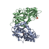
| ||||||||
|---|---|---|---|---|---|---|---|---|---|
| 1 |
| ||||||||
| Unit cell |
|
-Components
| #1: Protein | Mass: 28961.559 Da / Num. of mol.: 2 / Fragment: ligand binding domain Source method: isolated from a genetically manipulated source Details: The construct contains residues K409-E526 and T682-S815 of the ML032222a ligand binding domain connected by a synthetic GT linker Source: (gene. exp.) Mnemiopsis leidyi (sea walnut) / Gene: ML032222a / Plasmid: pET22 / Production host:  #2: Chemical |   Type: peptide linking / Mass: 75.067 Da / Num. of mol.: 2 / Source method: obtained synthetically / Formula: C2H5NO2 Type: peptide linking / Mass: 75.067 Da / Num. of mol.: 2 / Source method: obtained synthetically / Formula: C2H5NO2#3: Chemical | ChemComp-MG / |   Mass: 24.305 Da / Num. of mol.: 1 / Source method: obtained synthetically / Formula: Mg Mass: 24.305 Da / Num. of mol.: 1 / Source method: obtained synthetically / Formula: Mg#4: Chemical | ChemComp-SO4 / |   Mass: 96.063 Da / Num. of mol.: 1 / Source method: obtained synthetically / Formula: SO4 Mass: 96.063 Da / Num. of mol.: 1 / Source method: obtained synthetically / Formula: SO4#5: Water | ChemComp-HOH / |  Mass: 18.015 Da / Num. of mol.: 817 / Source method: isolated from a natural source / Formula: H2O Mass: 18.015 Da / Num. of mol.: 817 / Source method: isolated from a natural source / Formula: H2OHas protein modification | Y | Sequence details | The construct contains residues K409-E526 and T682-S815 of the ML032222a ligand binding domain ...The construct contains residues K409-E526 and T682-S815 of the ML032222a ligand binding domain connected by a synthetic GT linker | |
|---|
-Experimental details
-Experiment
| Experiment | Method: X-RAY DIFFRACTION / Number of used crystals: 1 |
|---|
- Sample preparation
| Crystal | Density Matthews: 2.47 Å3/Da / Density % sol: 50.24 % |
|---|---|
| Crystal grow | Temperature: 277 K / Method: vapor diffusion, hanging drop / pH: 6.5 Details: Reservoir: 18% PEG 8K, 100 mM Cacodylate, 150 mM MgS04. Protein buffer: 100 mM NaCl, 10 mM HEPES, pH 7.0, 0.5 mM EDTA, 2 mM Na glutamate, no added glycine PH range: 6.5 |
-Data collection
| Diffraction | Mean temperature: 100 K |
|---|---|
| Diffraction source | Source: SYNCHROTRON / Site: APS  / Beamline: 22-ID / Wavelength: 1 Å / Beamline: 22-ID / Wavelength: 1 Å |
| Detector | Type: MARMOSAIC 300 mm CCD / Detector: CCD / Date: Oct 14, 2013 |
| Radiation | Protocol: SINGLE WAVELENGTH / Monochromatic (M) / Laue (L): M / Scattering type: x-ray |
| Radiation wavelength | Wavelength: 1 Å / Relative weight: 1 |
| Reflection | Resolution: 1.21→40 Å / Num. obs: 169396 / % possible obs: 90.8 % / Redundancy: 3.6 % / Rmerge(I) obs: 0.055 / Net I/σ(I): 20.8 |
| Reflection shell | Resolution: 1.21→1.23 Å / Redundancy: 2.9 % / Rmerge(I) obs: 0.456 / Mean I/σ(I) obs: 2 / % possible all: 64.8 |
- Processing
| Software |
| |||||||||||||||||||||||||||||||||||||||||||||||||||||||||||||||||||||||||||||||||||||||||||||||||||||||||||||||||||||||||||||||||||||||||||||||||||||||||||||||||||||||||||||||||||||||||||||||||||||||||||||||||||||||||
|---|---|---|---|---|---|---|---|---|---|---|---|---|---|---|---|---|---|---|---|---|---|---|---|---|---|---|---|---|---|---|---|---|---|---|---|---|---|---|---|---|---|---|---|---|---|---|---|---|---|---|---|---|---|---|---|---|---|---|---|---|---|---|---|---|---|---|---|---|---|---|---|---|---|---|---|---|---|---|---|---|---|---|---|---|---|---|---|---|---|---|---|---|---|---|---|---|---|---|---|---|---|---|---|---|---|---|---|---|---|---|---|---|---|---|---|---|---|---|---|---|---|---|---|---|---|---|---|---|---|---|---|---|---|---|---|---|---|---|---|---|---|---|---|---|---|---|---|---|---|---|---|---|---|---|---|---|---|---|---|---|---|---|---|---|---|---|---|---|---|---|---|---|---|---|---|---|---|---|---|---|---|---|---|---|---|---|---|---|---|---|---|---|---|---|---|---|---|---|---|---|---|---|---|---|---|---|---|---|---|---|---|---|---|---|---|---|---|---|
| Refinement | Method to determine structure: MOLECULAR REPLACEMENT Starting model: 1S50 Resolution: 1.21→31.871 Å / SU ML: 0.09 / Cross valid method: THROUGHOUT / σ(F): 0.39 / Phase error: 16.77 / Stereochemistry target values: ML
| |||||||||||||||||||||||||||||||||||||||||||||||||||||||||||||||||||||||||||||||||||||||||||||||||||||||||||||||||||||||||||||||||||||||||||||||||||||||||||||||||||||||||||||||||||||||||||||||||||||||||||||||||||||||||
| Solvent computation | Shrinkage radii: 0.9 Å / VDW probe radii: 1.11 Å / Solvent model: FLAT BULK SOLVENT MODEL | |||||||||||||||||||||||||||||||||||||||||||||||||||||||||||||||||||||||||||||||||||||||||||||||||||||||||||||||||||||||||||||||||||||||||||||||||||||||||||||||||||||||||||||||||||||||||||||||||||||||||||||||||||||||||
| Refinement step | Cycle: LAST / Resolution: 1.21→31.871 Å
| |||||||||||||||||||||||||||||||||||||||||||||||||||||||||||||||||||||||||||||||||||||||||||||||||||||||||||||||||||||||||||||||||||||||||||||||||||||||||||||||||||||||||||||||||||||||||||||||||||||||||||||||||||||||||
| Refine LS restraints |
| |||||||||||||||||||||||||||||||||||||||||||||||||||||||||||||||||||||||||||||||||||||||||||||||||||||||||||||||||||||||||||||||||||||||||||||||||||||||||||||||||||||||||||||||||||||||||||||||||||||||||||||||||||||||||
| LS refinement shell |
|
