 Movie
Movie Controller

[English] 日本語
 Yorodumi
- PDB-1hlq: CRYSTAL STRUCTURE OF RHODOFERAX FERMENTANS HIGH POTENTIAL IRON-SU... -
+ Open data

- Basic information
| Entry | Database: PDB / ID: 1hlq | ||||||
|---|---|---|---|---|---|---|---|
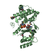
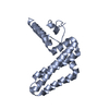
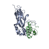
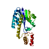
| Title | CRYSTAL STRUCTURE OF RHODOFERAX FERMENTANS HIGH POTENTIAL IRON-SULFUR PROTEIN REFINED TO 1.45 A | ||||||
 Components Components | HIGH-POTENTIAL IRON-SULFUR PROTEIN | ||||||
 Keywords Keywords | ELECTRON TRANSPORT / Iron sulfur cluster | ||||||
| Function / homology |  Function and homology information Function and homology informationaerobic electron transport chain / 4 iron, 4 sulfur cluster binding / electron transfer activity / metal ion binding Similarity search - Function | ||||||
| Biological species |  Rhodoferax fermentans (bacteria) Rhodoferax fermentans (bacteria) | ||||||
| Method |  X-RAY DIFFRACTION / SYNCHROTRON / MAD / Resolution: 1.45 Å X-RAY DIFFRACTION / SYNCHROTRON / MAD / Resolution: 1.45 Å | ||||||
 Authors Authors | Gonzalez, A. / Ciurli, S. / Benini, S. | ||||||
 Citation Citation | Journal: Acta Crystallogr.,Sect.D / Year: 2003 Title: Structure of Rhodoferax fermentans high-potential iron-sulfur protein solved by MAD. Authors: Gonzalez, A. / Benini, S. / Ciurli, S. #1: Journal: Eur.J.Biochem. / Year: 1997Title: The primary structure of Rhodoferax fermentans high potential iron-sulfur protein, an electron donor to the photosynthetic reaction center Authors: Van Driesche, G. / Ciurli, S. / Hochkoeppler, A. / Van Beeumen, J.J. #2: Journal: FEBS Lett. / Year: 1995Title: The high-potential iron-sulfur protein (HIPIP) from Rhodoferax fermentans is competent in photosynthetic electron transfer Authors: Hochkoeppler, A. / Ciurli, S. / Venturoli, G. / Zannoni, D. #3: Journal: Arch.Biochem.Biophys. / Year: 1995Title: Isolation, characterization, and functional role of the high potentialiron-sulfur protein (HIPIP) from Rhodopherax fermentans Authors: Hochkoeppler, A. / Kofod, P. / Ferro, G. / Ciurli, S. #4: Journal: Eur.J.Biochem. / Year: 1996Title: 1H NMR of high potential iron-sulfur protein from the purple non-sulfur bacterium Rhodoferax fermentans Authors: Ciurli, S. / Cremonini, M.A. / Kofod, P. / Luchinat, C. #5: Journal: Proc.Natl.Acad.Sci.USA / Year: 1996Title: Kinetics of photoinduced electron transfer from high potential iron-sulfur protein (HIPIP) to the photosynthetic reaction center of the purple phototroph Rhodoferax fermentans Authors: Hochkoeppler, A. / Zannoni, D. / Ciurli, S. / Meyer, T.E. / Cusanovich, M.A. / Tolin, G. | ||||||
| History |
|
- Structure visualization
| Structure viewer | Molecule: MolmilJmol/JSmol |
|---|
- Downloads & links
-Download
| PDBx/mmCIF format | 1hlq.cif.gz | 108.2 KB | Display | PDBx/mmCIF format |
|---|---|---|---|---|
| PDB format | pdb1hlq.ent.gz | 84.7 KB | Display | PDB format |
| PDBx/mmJSON format | 1hlq.json.gz | Tree view | PDBx/mmJSON format | |
| Others |  Other downloads Other downloads |
-Validation report
| Arichive directory | https://data.pdbj.org/pub/pdb/validation_reports/hl/1hlqftp://data.pdbj.org/pub/pdb/validation_reports/hl/1hlq | HTTPS FTP |
|---|
-Related structure data
| Similar structure data |
|---|


-Links
 PDBj
PDBj- Assembly
| Deposited unit | 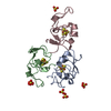
| ||||||||||
|---|---|---|---|---|---|---|---|---|---|---|---|
| 1 |
| ||||||||||
| Unit cell |
|
-Components
| #1: Protein | Mass: 7860.968 Da / Num. of mol.: 3 / Source method: isolated from a natural source / Source: (natural) Rhodoferax fermentans (bacteria) / References: UniProt: P80882#2: Chemical | ChemComp-SO4 /   Mass: 96.063 Da / Num. of mol.: 5 / Source method: obtained synthetically / Formula: SO4 Mass: 96.063 Da / Num. of mol.: 5 / Source method: obtained synthetically / Formula: SO4#3: Chemical |   Mass: 351.640 Da / Num. of mol.: 3 / Source method: obtained synthetically / Formula: Fe4S4 Mass: 351.640 Da / Num. of mol.: 3 / Source method: obtained synthetically / Formula: Fe4S4#4: Water | ChemComp-HOH / |  Mass: 18.015 Da / Num. of mol.: 260 / Source method: isolated from a natural source / Formula: H2O Mass: 18.015 Da / Num. of mol.: 260 / Source method: isolated from a natural source / Formula: H2O |
|---|
-Experimental details
-Experiment
| Experiment | Method: X-RAY DIFFRACTION / Number of used crystals: 2 |
|---|
- Sample preparation
| Crystal | Density Matthews: 2.11 Å3/Da / Density % sol: 41 % | ||||||||||||||||||||||||||||||||||||
|---|---|---|---|---|---|---|---|---|---|---|---|---|---|---|---|---|---|---|---|---|---|---|---|---|---|---|---|---|---|---|---|---|---|---|---|---|---|
| Crystal grow | Temperature: 290 K / Method: vapor diffusion, hanging drop / pH: 8 Details: HiPIP (20mg/ml, 20mM Tris pH 8, 2mM Beta-mercaptoethanol, 3.2M (NH4)2SO4 in 100mM Tris buffer, VAPOR DIFFUSION, HANGING DROP, temperature 290K | ||||||||||||||||||||||||||||||||||||
| Crystal grow | *PLUS Temperature: 293 K / Method: vapor diffusion | ||||||||||||||||||||||||||||||||||||
| Components of the solutions | *PLUS
|
-Data collection
| Diffraction |
| ||||||||||||||||||
|---|---|---|---|---|---|---|---|---|---|---|---|---|---|---|---|---|---|---|---|
| Diffraction source |
| ||||||||||||||||||
| Detector |
| ||||||||||||||||||
| Radiation |
| ||||||||||||||||||
| Radiation wavelength |
| ||||||||||||||||||
| Reflection | Resolution: 1.45→22.09 Å / Num. all: 42975 / Num. obs: 42975 / % possible obs: 99 % / Observed criterion σ(F): 0 / Observed criterion σ(I): 0 / Redundancy: 5.6 % / Biso Wilson estimate: 17.254 Å2 / Rmerge(I) obs: 0.047 / Net I/σ(I): 6.9 | ||||||||||||||||||
| Reflection shell | Resolution: 1.45→1.53 Å / Redundancy: 3.8 % / Rmerge(I) obs: 0.412 / Mean I/σ(I) obs: 0.6 / Num. unique all: 5912 / % possible all: 95.4 | ||||||||||||||||||
| Reflection | *PLUS Highest resolution: 1.45 Å / Lowest resolution: 22.09 Å / Num. obs: 42770 / % possible obs: 99 % / Num. measured all: 239390 | ||||||||||||||||||
| Reflection shell | *PLUS % possible obs: 95.4 % / Num. unique obs: 41858 / Num. measured obs: 238478 |

- Processing
| Software |
| ||||||||||||||||||||||||||||||||||||||||||||||||||||||||||||||||||||||||||||||||
|---|---|---|---|---|---|---|---|---|---|---|---|---|---|---|---|---|---|---|---|---|---|---|---|---|---|---|---|---|---|---|---|---|---|---|---|---|---|---|---|---|---|---|---|---|---|---|---|---|---|---|---|---|---|---|---|---|---|---|---|---|---|---|---|---|---|---|---|---|---|---|---|---|---|---|---|---|---|---|---|---|---|
| Refinement | Method to determine structure: MAD / Resolution: 1.45→22 Å / SU B: 0.83862 / SU ML: 0.03238 / Isotropic thermal model: Anisotropic / Cross valid method: FREE R-VALUE / σ(F): 0 / σ(I): 0 / ESU R: 0.07766 / Stereochemistry target values: Engh & Huber Details: Used conjugate direction method with maximum likelihood NCS and MAD phases were used as restrains
| ||||||||||||||||||||||||||||||||||||||||||||||||||||||||||||||||||||||||||||||||
| Refine analyze |
| ||||||||||||||||||||||||||||||||||||||||||||||||||||||||||||||||||||||||||||||||
| Refinement step | Cycle: LAST / Resolution: 1.45→22 Å
| ||||||||||||||||||||||||||||||||||||||||||||||||||||||||||||||||||||||||||||||||
| Refine LS restraints |
| ||||||||||||||||||||||||||||||||||||||||||||||||||||||||||||||||||||||||||||||||
| LS refinement shell | Resolution: 1.453→1.604 Å / Total num. of bins used: 10 /
| ||||||||||||||||||||||||||||||||||||||||||||||||||||||||||||||||||||||||||||||||
| Refinement | *PLUS | ||||||||||||||||||||||||||||||||||||||||||||||||||||||||||||||||||||||||||||||||
| Solvent computation | *PLUS | ||||||||||||||||||||||||||||||||||||||||||||||||||||||||||||||||||||||||||||||||
| Displacement parameters | *PLUS | ||||||||||||||||||||||||||||||||||||||||||||||||||||||||||||||||||||||||||||||||
| Refine LS restraints | *PLUS
|
