 Movie
Movie Controller


+ Open data

- Basic information
| Entry | Database: PDB / ID: 1hgz | ||||||
|---|---|---|---|---|---|---|---|
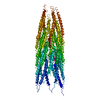
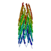
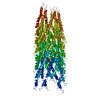
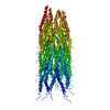
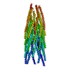
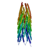
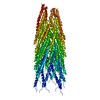
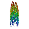
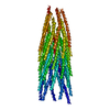
| Title | Filamentous Bacteriophage PH75 | ||||||
 Components Components | PH75 INOVIRUS MAJOR COAT PROTEIN | ||||||
 Keywords Keywords | VIRUS / VIRUS COAT PROTEIN / HELICAL VIRUS COAT PROTEIN / SSDNA VIRUSES / INOVIRUS / FILAMENTOUS BACTERIOPHAGE / THERMOPHILES / MEMBRANE PROTEINS / HELICAL VIRUS | ||||||
| Function / homology | Single alpha-helices involved in coiled-coils or other helix-helix interfaces - #80 / Bacteriophage M13, G8P, capsid domain superfamily / helical viral capsid / host cell membrane / Single alpha-helices involved in coiled-coils or other helix-helix interfaces / Up-down Bundle / Mainly Alpha / membrane / Capsid protein G8P Function and homology information Function and homology information | ||||||
| Biological species |   BACTERIOPHAGE PH75 (virus) BACTERIOPHAGE PH75 (virus) | ||||||
| Method |  FIBER DIFFRACTION / SYNCHROTRON / MOLECULAR REPLACEMENT / Resolution: 2.4 Å FIBER DIFFRACTION / SYNCHROTRON / MOLECULAR REPLACEMENT / Resolution: 2.4 Å | ||||||
 Authors Authors | Pederson, D.M. / Welsh, L.C. / Marvin, D.A. / Sampson, M. / Perham, R.N. / Yu, M. / Slater, M.R. | ||||||
 Citation Citation | Journal: J.Mol.Biol. / Year: 2001 Title: The Protein Capsid of Filamentous Bacteriophage Ph75 from Thermus Thermophilus Authors: Pederson, D.M. / Welsh, L.C. / Marvin, D.A. / Sampson, M. / Perham, R.N. / Yu, M. / Slater, M.R. #1: Journal: Acta Crystallogr.,Sect.D / Year: 2000Title: The Molecular Structure and Structural Transition of the Alpha-Helical Capsid in Filamentous Bacteriophage Pf1 Authors: Welsh, L.C. / Symmons, M.F. / Marvin, D.A. #2: Journal: J.Mol.Biol. / Year: 1998Title: Structure of the Capsid of Pf3 Filamentous Phage Determined from X-Ray Fibre Diffraction Data at 3.1 A Resolution Authors: Welsh, L.C. / Symmons, M.F. / Sturtevant, J.M. / Marvin, D.A. / Perham, R.N. #3: Journal: Acta Crystallogr.,Sect.D / Year: 1995Title: Pf1 Filamentous Bacteriophage: Refinement of a Molecular Model by Simulated Annealing Using 3.3 Angstroms Resolution X-Ray Fibre Diffraction Data Authors: Gonzalez, A. / Nave, C. / Marvin, D.A. #4: Journal: Int.J.Biol.Macromol. / Year: 1990Title: Model-Building Studies of Inovirus: Genetic Variations on a Geometric Theme Authors: Marvin, D.A. | ||||||
| History |
| ||||||
| Remark 650 | HELIX DETERMINATION METHOD: AUTHOR PROVIDED. |
- Structure visualization
| Structure viewer | Molecule: MolmilJmol/JSmol |
|---|
- Downloads & links
-Download
| PDBx/mmCIF format | 1hgz.cif.gz | 26.5 KB | Display | PDBx/mmCIF format |
|---|---|---|---|---|
| PDB format | pdb1hgz.ent.gz | 17.2 KB | Display | PDB format |
| PDBx/mmJSON format | 1hgz.json.gz | Tree view | PDBx/mmJSON format | |
| Others |  Other downloads Other downloads |
-Validation report
| Summary document | 1hgz_validation.pdf.gz | 342.7 KB | Display | wwPDB validaton report |
|---|---|---|---|---|
| Full document | 1hgz_full_validation.pdf.gz | 347.7 KB | Display | |
| Data in XML | 1hgz_validation.xml.gz | 3 KB | Display | |
| Data in CIF | 1hgz_validation.cif.gz | 3.8 KB | Display | |
| Arichive directory | https://data.pdbj.org/pub/pdb/validation_reports/hg/1hgzftp://data.pdbj.org/pub/pdb/validation_reports/hg/1hgz | HTTPS FTP |
-Related structure data
| Related structure data |  1hgvC  1hh0C 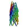 1ifnS S: Starting model for refinement C: citing same article ( |
|---|---|
| Similar structure data |


-Links
 PDBj
PDBj- Assembly
| Deposited unit | 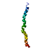
| ||||||||
|---|---|---|---|---|---|---|---|---|---|
| 1 | x 35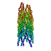
| ||||||||
| 2 |
| ||||||||
| 3 | 
| ||||||||
| Unit cell |
| ||||||||
| Symmetry | Helical symmetry: (Circular symmetry: 1 / Dyad axis: no / N subunits divisor: 1 / Num. of operations: 35 / Rise per n subunits: 2.9 Å / Rotation per n subunits: 66.667 °) |
-Components
| #1: Protein/peptide | Mass: 4816.578 Da / Num. of mol.: 1 Source method: isolated from a genetically manipulated source Details: MAJOR COAT PROTEIN ASSEMBLY / Source: (gene. exp.) BACTERIOPHAGE PH75 (virus) / Description: GROWN IN THERMUS THERMOPHILUS / Production host:  THERMUS THERMOPHILUS HB8 (bacteria) / References: UniProt: P82889 THERMUS THERMOPHILUS HB8 (bacteria) / References: UniProt: P82889 |
|---|
-Experimental details
-Experiment
| Experiment | Method: FIBER DIFFRACTION |
|---|
- Sample preparation
| Crystal | Description: THE DATA IS DERIVED FROM CONTINUOUS TRANSFORM DATA AND THEREFORE THE NUMBER OF UNIQUE REFLECTIONS IS A MEANINGLESS NUMBER. THE STRUCTURE WAS REFINED AGAI THE SAME STRUCTURE FACTORS AS ...Description: THE DATA IS DERIVED FROM CONTINUOUS TRANSFORM DATA AND THEREFORE THE NUMBER OF UNIQUE REFLECTIONS IS A MEANINGLESS NUMBER. THE STRUCTURE WAS REFINED AGAI THE SAME STRUCTURE FACTORS AS PDB ENTRY 1HGV, USING DIFFERENT REFINEMENT PROTOCOL. THEREFORE, THE STRUCT FACTORS FOR 1HGZ, CAN BE TAKEN FROM R1HGVSF |
|---|---|
| Crystal grow | pH: 7.5 / Details: pH 7.50 |
| Crystal grow | *PLUS Method: unknown |
-Data collection
| Diffraction | Mean temperature: 300 K |
|---|---|
| Diffraction source | Source: SYNCHROTRON / Site: SRS  / Beamline: PX7.2 / Wavelength: 1.488 / Beamline: PX7.2 / Wavelength: 1.488 |
| Detector | Type: MARRESEARCH / Detector: IMAGE PLATE / Date: Feb 15, 1999 / Details: MIRRORS |
| Radiation | Monochromator: GE CRYSTAL / Protocol: SINGLE WAVELENGTH / Monochromatic (M) / Laue (L): M / Scattering type: x-ray |
| Radiation wavelength | Wavelength: 1.488 Å / Relative weight: 1 |
| Reflection | Resolution: 2.4→50 Å / % possible obs: 100 % |
| Reflection | *PLUS % possible obs: 100 % |
- Processing
| Software |
| ||||||||||||||||||||||||||||||||||||||||||||||||||||||||||||
|---|---|---|---|---|---|---|---|---|---|---|---|---|---|---|---|---|---|---|---|---|---|---|---|---|---|---|---|---|---|---|---|---|---|---|---|---|---|---|---|---|---|---|---|---|---|---|---|---|---|---|---|---|---|---|---|---|---|---|---|---|---|
| Refinement | Method to determine structure: MOLECULAR REPLACEMENT Starting model: 1IFN Resolution: 2.4→50 Å / σ(F): 0 Details: THE MODEL WAS DERIVED FROM PDB ENTRY 1IFN, REFERENCE 1, BY MOLECULAR REPLACEMENT
| ||||||||||||||||||||||||||||||||||||||||||||||||||||||||||||
| Refinement step | Cycle: LAST / Resolution: 2.4→50 Å
| ||||||||||||||||||||||||||||||||||||||||||||||||||||||||||||
| Refine LS restraints |
| ||||||||||||||||||||||||||||||||||||||||||||||||||||||||||||
| Software | *PLUS Name: FXPLOR / Classification: refinement | ||||||||||||||||||||||||||||||||||||||||||||||||||||||||||||
| Refinement | *PLUS Rfactor obs: 0.22 / Rfactor Rfree: 0.4 | ||||||||||||||||||||||||||||||||||||||||||||||||||||||||||||
| Solvent computation | *PLUS | ||||||||||||||||||||||||||||||||||||||||||||||||||||||||||||
| Displacement parameters | *PLUS | ||||||||||||||||||||||||||||||||||||||||||||||||||||||||||||
| Refine LS restraints | *PLUS
|
