 Movie
Movie Controller

[English] 日本語
 Yorodumi
- PDB-4ifm: PF1 FILAMENTOUS BACTERIOPHAGE: REFINEMENT OF A MOLECULAR MODEL BY... -
+ Open data

- Basic information
| Entry | Database: PDB / ID: 4ifm | ||||||
|---|---|---|---|---|---|---|---|
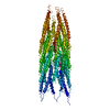
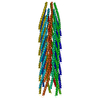
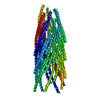
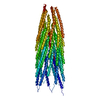
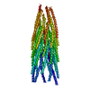
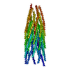
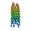
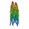
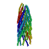
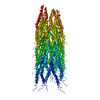
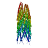
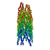
| Title | PF1 FILAMENTOUS BACTERIOPHAGE: REFINEMENT OF A MOLECULAR MODEL BY SIMULATED ANNEALING USING 3.3 ANGSTROMS RESOLUTION X-RAY FIBRE DIFFRACTION DATA | ||||||
 Components Components | PF1 FILAMENTOUS BACTERIOPHAGE | ||||||
 Keywords Keywords | VIRUS / VIRUS COAT PROTEIN / Helical virus | ||||||
| Function / homology | Single alpha-helices involved in coiled-coils or other helix-helix interfaces - #230 / Inovirus Coat protein B / Capsid protein G8P / helical viral capsid / host cell membrane / Single alpha-helices involved in coiled-coils or other helix-helix interfaces / Up-down Bundle / Mainly Alpha / Capsid protein G8P Function and homology information Function and homology information | ||||||
| Biological species |  Pseudomonas phage Pf1 (virus) Pseudomonas phage Pf1 (virus) | ||||||
| Method |  FIBER DIFFRACTION / Resolution: 3.3 Å FIBER DIFFRACTION / Resolution: 3.3 Å | ||||||
 Authors Authors | Marvin, D.A. | ||||||
 Citation Citation | Journal: Acta Crystallogr.,Sect.D / Year: 1995 Title: Pf1 filamentous bacteriophage: refinement of a molecular model by simulated annealing using 3.3 A resolution X-ray fibre diffraction data. Authors: Gonzalez, A. / Nave, C. / Marvin, D.A. #1: Journal: Phase Transitions / Year: 1992Title: Two Forms of Pf1 Inovirus: X-Ray Diffraction Studies on a Structural Phase Transition and a Calculated Libration Normal Mode of the Asymmetric Unit Authors: Marvin, D.A. / Nave, C. / Bansal, M. / Hale, R.D. / Salje, E.K.H. #2: Journal: Int.J.Biol.Macromol. / Year: 1990Title: Model-Building Studies of Inovirus: Genetic Variations on a Geometric Theme Authors: Marvin, D.A. #3: Journal: Int.J.Biol.Macromol. / Year: 1989Title: Dynamics of Telescoping Inovirus: A Mechanism for Assembly at Membrane Adhesions Authors: Marvin, D.A. | ||||||
| History |
|
- Structure visualization
| Structure viewer | Molecule: MolmilJmol/JSmol |
|---|
- Downloads & links
-Download
| PDBx/mmCIF format | 4ifm.cif.gz | 16.4 KB | Display | PDBx/mmCIF format |
|---|---|---|---|---|
| PDB format | pdb4ifm.ent.gz | 10.5 KB | Display | PDB format |
| PDBx/mmJSON format | 4ifm.json.gz | Tree view | PDBx/mmJSON format | |
| Others |  Other downloads Other downloads |
-Validation report
| Arichive directory | https://data.pdbj.org/pub/pdb/validation_reports/if/4ifmftp://data.pdbj.org/pub/pdb/validation_reports/if/4ifm | HTTPS FTP |
|---|
-Related structure data


-Links
 PDBj
PDBj- Assembly
| Deposited unit | 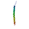
| ||||||||
|---|---|---|---|---|---|---|---|---|---|
| 1 | x 35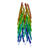
| ||||||||
| 2 |
| ||||||||
| 3 | 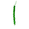
| ||||||||
| Unit cell |
| ||||||||
| Symmetry | Helical symmetry: (Circular symmetry: 1 / Dyad axis: no / N subunits divisor: 1 / Num. of operations: 35 / Rise per n subunits: 3.05 Å / Rotation per n subunits: 65.915 °) |
-Components
| #1: Protein/peptide | Mass: 4612.393 Da / Num. of mol.: 1 / Source method: isolated from a natural source / Details: GROWN IN PSEUDOMONAS AERUGINOSA / Source: (natural) Pseudomonas phage Pf1 (virus) / Genus: Inovirus / References: UniProt: P03621 |
|---|
-Experimental details
-Experiment
| Experiment | Method: FIBER DIFFRACTION |
|---|
- Processing
| Software |
| ||||||||||||
|---|---|---|---|---|---|---|---|---|---|---|---|---|---|
| Refinement | Highest resolution: 3.3 Å Details: COORDINATES ARE GIVEN FOR A SINGLE ASYMMETRIC UNIT OF THE COAT PROTEIN ASSEMBLY. THE COMPLETE PROTEIN ASSEMBLY CONTAINS SEVERAL THOUSAND ASYMMETRIC UNITS; THE EXACT NUMBER DEPENDS ON THE ...Details: COORDINATES ARE GIVEN FOR A SINGLE ASYMMETRIC UNIT OF THE COAT PROTEIN ASSEMBLY. THE COMPLETE PROTEIN ASSEMBLY CONTAINS SEVERAL THOUSAND ASYMMETRIC UNITS; THE EXACT NUMBER DEPENDS ON THE LENGTH OF THE DNA. THE PROTEIN ASSEMBLY FORMS A CYLINDRICAL SHELL SURROUNDING A DNA CORE. | ||||||||||||
| Refinement step | Cycle: LAST / Highest resolution: 3.3 Å
|
