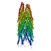
 Movie
Movie Controller


+ Open data

- Basic information
| Entry | Database: PDB / ID: 1hgv | ||||||
|---|---|---|---|---|---|---|---|
| Title | Filamentous Bacteriophage PH75 | ||||||
 Components Components | PH75 INOVIRUS MAJOR COAT PROTEIN | ||||||
 Keywords Keywords | VIRUS / VIRUS COAT PROTEIN / HELICAL VIRUS COAT PROTEIN / SSDNA VIRUSES / INOVIRUS / FILAMENTOUS BACTERIOPHAGE / THERMOPHILES / MEMBRANE PROTEINS / ICOSAHEDRAL VIRUS | ||||||
| Function / homology | Single alpha-helices involved in coiled-coils or other helix-helix interfaces - #80 / Bacteriophage M13, G8P, capsid domain superfamily / helical viral capsid / host cell membrane / Single alpha-helices involved in coiled-coils or other helix-helix interfaces / Up-down Bundle / Mainly Alpha / Capsid protein G8P Function and homology information Function and homology information | ||||||
| Biological species |   BACTERIOPHAGE PH75 (virus) BACTERIOPHAGE PH75 (virus) | ||||||
| Method |  FIBER DIFFRACTION / SYNCHROTRON / MOLECULAR REPLACEMENT / Resolution: 2.4 Å FIBER DIFFRACTION / SYNCHROTRON / MOLECULAR REPLACEMENT / Resolution: 2.4 Å | ||||||
 Authors Authors | Pederson, D.M. / Welsh, L.C. / Marvin, D.A. / Sampson, M. / Perham, R.N. / Yu, M. / Slater, M.R. | ||||||
 Citation Citation | Journal: J.Mol.Biol. / Year: 2001 Title: The Protein Capsid of Filamentous Bacteriophage Ph75 from Thermus Thermophilus Authors: Pederson, D.M. / Welsh, L.C. / Marvin, D.A. / Sampson, M. / Perham, R.N. / Yu, M. / Slater, M.R. #1: Journal: Acta Crystallogr.,Sect.D / Year: 2000Title: The Molecular Structure and Structural Transition of the Alpha-Helical Capsid in Filamentous Bacteriophage Pf1 Authors: Welsh, L.C. / Symmons, M.F. / Marvin, D.A. #2: Journal: J.Mol.Biol. / Year: 1998Title: Structure of the Capsid of Pf3 Filamentous Phage Determined from X-Ray Fibre Diffraction Data at 3.1 A Resolution Authors: Welsh, L.C. / Symmons, M.F. / Sturtevant, J.M. / Marvin, D.A. / Perham, R.N. #3: Journal: Acta Crystallogr.,Sect.D / Year: 1995Title: Pf1 Filamentous Bacteriophage: Refinement of a Molecular Model by Simulated Annealing Using 3.3 Angstroms Resolution X-Ray Fibre Diffraction Data Authors: Gonzalez, A. / Nave, C. / Marvin, D.A. #4: Journal: Int.J.Biol.Macromol. / Year: 1990Title: Model-Building Studies of Inovirus: Genetic Variations on a Geometric Theme Authors: Marvin, D.A. | ||||||
| History |
| ||||||
| Remark 285 | THE ANALOGUE OF THE CRYSTALLOGRAPHIC SPACE GROUP FOR HELICAL STRUCTURES IS THE LINE GROUP (A.KLUG, ... THE ANALOGUE OF THE CRYSTALLOGRAPHIC SPACE GROUP FOR HELICAL STRUCTURES IS THE LINE GROUP (A.KLUG, F.H.C.CRICK, H.W.WYCKOFF, ACTA CRYSTALLOG. V.11, 199, 1958). THE LINE GROUP OF PF1 IS S. THE UNIT CELL DIMENSIONS ARE THE HELIX PARAMETERS (UNIT TWIST TAU, UNIT HEIGHT P). THE INDEXING OF UNITS ALONG THE BASIC HELIX IS ILLUSTRATED IN REFERENCE 4. TO GENERATE COORDINATES X(K), Y(K), Z(K) OF UNIT K FROM THE GIVEN COORDINATES X(0), Y(0), Z(0) OF UNIT 0 IN A UNIT CELL WITH HELIX PARAMETERS (TAU, P) = (66.667, 2.90), APPLY THE MATRIX AND VECTOR: | COS(TAU*K) -SIN(TAU*K) 0 | | 0 | | SIN(TAU*K) COS(TAU*K) 0 | + | 0 | | 0 0 1 | | P*K | THE NEIGHBORS IN CONTACT WITH UNIT 0 ARE UNITS K = +/-1, +/-5, +/-6, +/-11 AND +/-17. THESE SYMMETRY-RELATED COPIES ARE USED TO DETERMINE INTERCHAIN NON-BONDED CONTACTS DURING THE REFINEMENT. |
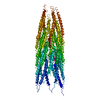
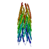
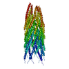
- Structure visualization
| Structure viewer | Molecule: MolmilJmol/JSmol |
|---|
- Downloads & links
-Download
| PDBx/mmCIF format | 1hgv.cif.gz | 27.1 KB | Display | PDBx/mmCIF format |
|---|---|---|---|---|
| PDB format | pdb1hgv.ent.gz | 17.8 KB | Display | PDB format |
| PDBx/mmJSON format | 1hgv.json.gz | Tree view | PDBx/mmJSON format | |
| Others |  Other downloads Other downloads |
-Validation report
| Arichive directory | https://data.pdbj.org/pub/pdb/validation_reports/hg/1hgvftp://data.pdbj.org/pub/pdb/validation_reports/hg/1hgv | HTTPS FTP |
|---|
-Related structure data
| Related structure data |  1hgzC  1hh0C 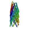 1ifnS C: citing same article ( S: Starting model for refinement |
|---|---|
| Similar structure data |


-Links
 PDBj
PDBj- Assembly
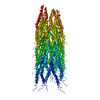
| Deposited unit | 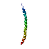
| ||||||||
|---|---|---|---|---|---|---|---|---|---|
| 1 | x 35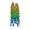
| ||||||||
| 2 |
| ||||||||
| 3 | 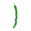
| ||||||||
| Unit cell |
| ||||||||
| Symmetry | Helical symmetry: (Circular symmetry: 1 / Dyad axis: no / N subunits divisor: 1 / Num. of operations: 35 / Rise per n subunits: 2.9 Å / Rotation per n subunits: 66.667 °) |
-Components
| #1: Protein/peptide | Mass: 4816.578 Da / Num. of mol.: 1 Source method: isolated from a genetically manipulated source Details: MAJOR COAT PROTEIN ASSEMBLY / Source: (gene. exp.) BACTERIOPHAGE PH75 (virus) / Description: GROWN IN THERMUS THERMOPHILUS / Production host:  THERMUS THERMOPHILUS HB8 (bacteria) / References: UniProt: P82889 THERMUS THERMOPHILUS HB8 (bacteria) / References: UniProt: P82889 |
|---|
-Experimental details
-Experiment
| Experiment | Method: FIBER DIFFRACTION |
|---|
- Sample preparation
| Crystal | Description: THE DATA IS DERIVED FROM CONTINUOUS TRANSFORM DATA AND THEREFORE THE NUMBER OF UNIQUE REFLECTIONS IS A MEANINGLESS NUMBER. |
|---|---|
| Crystal grow | pH: 7.5 / Details: pH 7.50 |
| Crystal grow | *PLUS Method: unknown |
-Data collection
| Diffraction | Mean temperature: 300 K |
|---|---|
| Diffraction source | Source: SYNCHROTRON / Site: SRS  / Beamline: PX7.2 / Wavelength: 1.488 / Beamline: PX7.2 / Wavelength: 1.488 |
| Detector | Type: MARRESEARCH / Detector: IMAGE PLATE / Date: Feb 15, 1999 / Details: MIRRORS |
| Radiation | Monochromator: GE CRYSTAL / Protocol: SINGLE WAVELENGTH / Monochromatic (M) / Laue (L): M / Scattering type: x-ray |
| Radiation wavelength | Wavelength: 1.488 Å / Relative weight: 1 |
| Reflection | Resolution: 2.4→50 Å / % possible obs: 100 % |
| Reflection | *PLUS % possible obs: 100 % |
- Processing
| Software |
| ||||||||||||||||||||||||||||||||||||||||||||||||||||||||||||
|---|---|---|---|---|---|---|---|---|---|---|---|---|---|---|---|---|---|---|---|---|---|---|---|---|---|---|---|---|---|---|---|---|---|---|---|---|---|---|---|---|---|---|---|---|---|---|---|---|---|---|---|---|---|---|---|---|---|---|---|---|---|
| Refinement | Method to determine structure: MOLECULAR REPLACEMENT Starting model: PDB ENTRY 1IFN Resolution: 2.4→50 Å / σ(F): 0 Details: THE MODEL WAS DERIVED FROM PDB ENTRY 1IFN, REFERENCE 1, BY MOLECULAR REPLACEMENT
| ||||||||||||||||||||||||||||||||||||||||||||||||||||||||||||
| Refinement step | Cycle: LAST / Resolution: 2.4→50 Å
| ||||||||||||||||||||||||||||||||||||||||||||||||||||||||||||
| Refine LS restraints |
| ||||||||||||||||||||||||||||||||||||||||||||||||||||||||||||
| Software | *PLUS Name: FXPLOR / Classification: refinement | ||||||||||||||||||||||||||||||||||||||||||||||||||||||||||||
| Refinement | *PLUS Rfactor obs: 0.19 / Rfactor Rfree: 0.4 | ||||||||||||||||||||||||||||||||||||||||||||||||||||||||||||
| Solvent computation | *PLUS | ||||||||||||||||||||||||||||||||||||||||||||||||||||||||||||
| Displacement parameters | *PLUS | ||||||||||||||||||||||||||||||||||||||||||||||||||||||||||||
| Refine LS restraints | *PLUS
|
