 Movie
Movie Controller

[English] 日本語
 Yorodumi
- PDB-1h4r: Crystal Structure of the FERM domain of Merlin, the Neurofibromat... -
+ Open data

- Basic information
| Entry | Database: PDB / ID: 1h4r | ||||||
|---|---|---|---|---|---|---|---|
| Title | Crystal Structure of the FERM domain of Merlin, the Neurofibromatosis 2 Tumor Suppressor Protein. | ||||||
 Components Components | MERLIN | ||||||
 Keywords Keywords | STRUCTURAL PROTEIN / FERM / NEUROFIBROMATOSIS / NF2 / CYTOSKELETON / ANTI-ONCOGENE | ||||||
| Function / homology |  Function and homology information Function and homology informationregulation of hippo signaling / negative regulation of cell growth involved in contact inhibition / regulation of organelle assembly / regulation of gliogenesis / positive regulation of early endosome to late endosome transport / Schwann cell proliferation / osteoblast proliferation / negative regulation of Schwann cell proliferation / negative regulation of osteoblast proliferation / positive regulation of protein localization to early endosome ...regulation of hippo signaling / negative regulation of cell growth involved in contact inhibition / regulation of organelle assembly / regulation of gliogenesis / positive regulation of early endosome to late endosome transport / Schwann cell proliferation / osteoblast proliferation / negative regulation of Schwann cell proliferation / negative regulation of osteoblast proliferation / positive regulation of protein localization to early endosome / ectoderm development / lens fiber cell differentiation / regulation of stem cell proliferation / regulation of neural precursor cell proliferation / hippo signaling / negative regulation of receptor signaling pathway via JAK-STAT / cell-cell junction organization / regulation of protein localization to nucleus / filopodium membrane / negative regulation of cell-cell adhesion / cortical actin cytoskeleton / odontogenesis of dentin-containing tooth / cleavage furrow / RHO GTPases activate PAKs / mesoderm formation / negative regulation of cell-matrix adhesion / positive regulation of stress fiber assembly / negative regulation of MAPK cascade / signaling adaptor activity / negative regulation of cell migration / filopodium / hippocampus development / adherens junction / positive regulation of cell differentiation / regulation of protein stability / Regulation of actin dynamics for phagocytic cup formation / integrin binding / ruffle membrane / apical part of cell / regulation of cell shape / MAPK cascade / lamellipodium / actin binding / actin cytoskeleton organization / cell body / regulation of apoptotic process / cytoskeleton / early endosome / regulation of cell cycle / neuron projection / negative regulation of cell population proliferation / nucleolus / perinuclear region of cytoplasm / membrane / nucleus / plasma membrane / cytoplasm / cytosol Similarity search - Function | ||||||
| Biological species |  HOMO SAPIENS (human) HOMO SAPIENS (human) | ||||||
| Method |  X-RAY DIFFRACTION / SYNCHROTRON / MOLECULAR REPLACEMENT / Resolution: 1.8 Å X-RAY DIFFRACTION / SYNCHROTRON / MOLECULAR REPLACEMENT / Resolution: 1.8 Å | ||||||
 Authors Authors | Cooper, D.R. / Kang, B.S. / Sheffield, P. / Devedjiev, Y. / Derewenda, Z.S. | ||||||
 Citation Citation | Journal: Acta Crystallogr.,Sect.D / Year: 2002 Title: The Structure of the Ferm Domain of Merlin, the Neurofibromatosis Type 2 Gene Product. Authors: Kang, B.S. / Cooper, D.R. / Devedjiev, Y. / Derewenda, U. / Derewenda, Z.S. | ||||||
| History |
|
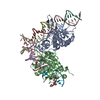
- Structure visualization
| Structure viewer | Molecule: MolmilJmol/JSmol |
|---|
- Downloads & links
-Download
| PDBx/mmCIF format | 1h4r.cif.gz | 151.4 KB | Display | PDBx/mmCIF format |
|---|---|---|---|---|
| PDB format | pdb1h4r.ent.gz | 120.2 KB | Display | PDB format |
| PDBx/mmJSON format | 1h4r.json.gz | Tree view | PDBx/mmJSON format | |
| Others |  Other downloads Other downloads |
-Validation report
| Arichive directory | https://data.pdbj.org/pub/pdb/validation_reports/h4/1h4rftp://data.pdbj.org/pub/pdb/validation_reports/h4/1h4r | HTTPS FTP |
|---|
-Related structure data
| Related structure data |  1gc6S S: Starting model for refinement |
|---|---|
| Similar structure data |


-Links
 PDBj
PDBj


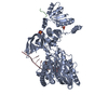
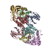
- Assembly
| Deposited unit | 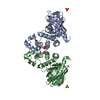
| ||||||||
|---|---|---|---|---|---|---|---|---|---|
| 1 |
| ||||||||
| Unit cell |
|
-Components
| #1: Protein | Mass: 36987.848 Da / Num. of mol.: 2 / Fragment: FERM DOMAIN RESIDUES 1-313 Source method: isolated from a genetically manipulated source Source: (gene. exp.) HOMO SAPIENS (human)Description: RECOMBINANTLY EXPRESSED IN BL21-RIL CELLS AS A HEXA-HISTIDINE AND GST TAGGED PROTEIN. THE TAG WAS REMOVED BY RTEV CLEAVAGE. Plasmid: PHGM313 / Production host:  #2: Chemical | ChemComp-SO4 /   Mass: 96.063 Da / Num. of mol.: 6 / Source method: obtained synthetically / Formula: SO4 Mass: 96.063 Da / Num. of mol.: 6 / Source method: obtained synthetically / Formula: SO4#3: Water | ChemComp-HOH / |  Mass: 18.015 Da / Num. of mol.: 861 / Source method: isolated from a natural source / Formula: H2O Mass: 18.015 Da / Num. of mol.: 861 / Source method: isolated from a natural source / Formula: H2OCompound details | ACTS AS A MEMBRANE STABILIZIN | Sequence details | N-TERMINAL GLYCINE IS FROM RTEV CLEAVAGE. | |
|---|
-Experimental details
-Experiment
| Experiment | Method: X-RAY DIFFRACTION / Number of used crystals: 1 |
|---|
- Sample preparation
| Crystal | Density Matthews: 2.5 Å3/Da / Density % sol: 51.4 % | |||||||||||||||||||||||||
|---|---|---|---|---|---|---|---|---|---|---|---|---|---|---|---|---|---|---|---|---|---|---|---|---|---|---|
| Crystal grow | Method: vapor diffusion, hanging drop / pH: 6.5 Details: THE PROTEIN WAS CRYSTALLIZED USING HANGING-DROP VAPOR DIFFUSION WIHTH 56% AMMONIUM SULFATE, 2% DIOXANE, 100 MM CACODYLATE, PH 6.5. A 1:1 RATIO OF PROTEIN TO WELL SOLUTION WAS USED. | |||||||||||||||||||||||||
| Crystal grow | *PLUS Temperature: 294 K / Method: vapor diffusion, sitting drop | |||||||||||||||||||||||||
| Components of the solutions | *PLUS
|
-Data collection
| Diffraction | Mean temperature: 100 K |
|---|---|
| Diffraction source | Source: SYNCHROTRON / Site: NSLS  / Beamline: X9B / Wavelength: 0.92 / Beamline: X9B / Wavelength: 0.92 |
| Detector | Type: ADSC CCD / Detector: CCD / Date: Mar 15, 2001 |
| Radiation | Protocol: SINGLE WAVELENGTH / Monochromatic (M) / Laue (L): M / Scattering type: x-ray |
| Radiation wavelength | Wavelength: 0.92 Å / Relative weight: 1 |
| Reflection | Resolution: 1.8→25 Å / Num. obs: 68182 / % possible obs: 95.5 % / Redundancy: 3.5 % / Rmerge(I) obs: 0.065 / Net I/σ(I): 16.8 |
| Reflection shell | Resolution: 1.8→1.86 Å / Redundancy: 3.4 % / Rmerge(I) obs: 0.662 / Mean I/σ(I) obs: 2.33 / % possible all: 97.2 |
| Reflection | *PLUS Highest resolution: 1.8 Å / Lowest resolution: 30 Å / Num. obs: 68222 / % possible obs: 95.4 % / Redundancy: 3.6 % |
| Reflection shell | *PLUS % possible obs: 97.2 % / Num. unique obs: 6875 / Rmerge(I) obs: 0.622 |
- Processing
| Software |
| ||||||||||||||||||||||||||||||||||||||||||||||||||||||||||||||||||||||||||||||||||||||||||||||||||||||||||||||||||||||||||||||||||||||||||||||||||||||||||||||||||||||||||||||||||||||
|---|---|---|---|---|---|---|---|---|---|---|---|---|---|---|---|---|---|---|---|---|---|---|---|---|---|---|---|---|---|---|---|---|---|---|---|---|---|---|---|---|---|---|---|---|---|---|---|---|---|---|---|---|---|---|---|---|---|---|---|---|---|---|---|---|---|---|---|---|---|---|---|---|---|---|---|---|---|---|---|---|---|---|---|---|---|---|---|---|---|---|---|---|---|---|---|---|---|---|---|---|---|---|---|---|---|---|---|---|---|---|---|---|---|---|---|---|---|---|---|---|---|---|---|---|---|---|---|---|---|---|---|---|---|---|---|---|---|---|---|---|---|---|---|---|---|---|---|---|---|---|---|---|---|---|---|---|---|---|---|---|---|---|---|---|---|---|---|---|---|---|---|---|---|---|---|---|---|---|---|---|---|---|---|
| Refinement | Method to determine structure: MOLECULAR REPLACEMENT Starting model: PDB ENTRY 1GC6.PDB Resolution: 1.8→25 Å / Cor.coef. Fo:Fc: 0.96 / Cor.coef. Fo:Fc free: 0.943 / SU B: 3.675 / SU ML: 0.116 / Cross valid method: THROUGHOUT / ESU R: 0.13 / ESU R Free: 0.123 / Stereochemistry target values: MAXIMUM LIKELIHOOD / Details: HYDROGENS HAVE BEEN ADDED IN THE RIDING POSITIONS
| ||||||||||||||||||||||||||||||||||||||||||||||||||||||||||||||||||||||||||||||||||||||||||||||||||||||||||||||||||||||||||||||||||||||||||||||||||||||||||||||||||||||||||||||||||||||
| Solvent computation | Ion probe radii: 0.8 Å / Shrinkage radii: 0.8 Å / VDW probe radii: 1.4 Å / Solvent model: BABINET MODEL PLUS MASK | ||||||||||||||||||||||||||||||||||||||||||||||||||||||||||||||||||||||||||||||||||||||||||||||||||||||||||||||||||||||||||||||||||||||||||||||||||||||||||||||||||||||||||||||||||||||
| Refinement step | Cycle: LAST / Resolution: 1.8→25 Å
| ||||||||||||||||||||||||||||||||||||||||||||||||||||||||||||||||||||||||||||||||||||||||||||||||||||||||||||||||||||||||||||||||||||||||||||||||||||||||||||||||||||||||||||||||||||||
| Refine LS restraints |
|