 Movie
Movie Controller

[English] 日本語
 Yorodumi
- PDB-1h4d: Biochemical and Structural Analysis of the Molybdenum Cofactor Bi... -
+ Open data

- Basic information
| Entry | Database: PDB / ID: 1h4d | ||||||
|---|---|---|---|---|---|---|---|
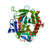
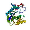
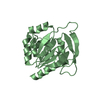
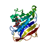
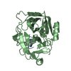
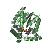
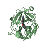
| Title | Biochemical and Structural Analysis of the Molybdenum Cofactor Biosynthesis protein MobA | ||||||
 Components Components | MOLYBDOPTERIN-GUANINE DINUCLEOTIDE BIOSYNTHESIS PROTEIN A | ||||||
 Keywords Keywords | MOLYBDENUM COFACTOR BIOSYNTHESIS / MOLYBDOPTERIN NUCLEOTIDYL-TRANSFERASE / GTP-BINDING | ||||||
| Function / homology |  Function and homology information Function and homology informationbis(molybdopterin guanine dinucleotide)molybdenum biosynthetic process / molybdenum cofactor guanylyltransferase / molybdenum cofactor guanylyltransferase activity / nucleotidyltransferase activity / GTP binding / magnesium ion binding / cytoplasm Similarity search - Function | ||||||
| Biological species |  | ||||||
| Method |  X-RAY DIFFRACTION / SYNCHROTRON / MOLECULAR REPLACEMENT / Resolution: 1.74 Å X-RAY DIFFRACTION / SYNCHROTRON / MOLECULAR REPLACEMENT / Resolution: 1.74 Å | ||||||
 Authors Authors | Guse, A. / Stevenson, C.E.M. / Kuper, J. / Buchanan, G. / Schwarz, G. / Mendel, R.R. / Lawson, D.M. / Palmer, T. | ||||||
 Citation Citation | Journal: J.Biol.Chem. / Year: 2003 Title: Biochemical and Structural Analysis of the Molybdenum Cofactor Biosynthesis Protein Moba Authors: Guse, A. / Stevenson, C.E.M. / Kuper, J. / Buchanan, G. / Schwarz, G. / Giordano, G. / Magalon, A. / Mendel, R.R. / Lawson, D.M. / Palmer, T. | ||||||
| History |
|
- Structure visualization
| Structure viewer | Molecule: MolmilJmol/JSmol |
|---|
- Downloads & links
-Download
| PDBx/mmCIF format | 1h4d.cif.gz | 57.2 KB | Display | PDBx/mmCIF format |
|---|---|---|---|---|
| PDB format | pdb1h4d.ent.gz | 39.8 KB | Display | PDB format |
| PDBx/mmJSON format | 1h4d.json.gz | Tree view | PDBx/mmJSON format | |
| Others |  Other downloads Other downloads |
-Validation report
| Arichive directory | https://data.pdbj.org/pub/pdb/validation_reports/h4/1h4dftp://data.pdbj.org/pub/pdb/validation_reports/h4/1h4d | HTTPS FTP |
|---|
-Related structure data
| Related structure data |  1h4cC  1h4eC  1hjjC  1hjlC  1e5kS S: Starting model for refinement C: citing same article ( |
|---|---|
| Similar structure data |

-Links
 PDBj
PDBj


- Assembly
| Deposited unit | 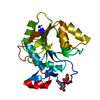
| ||||||||
|---|---|---|---|---|---|---|---|---|---|
| 1 |
| ||||||||
| Unit cell |
| ||||||||
| Components on special symmetry positions |
|
-Components
| #1: Protein | Mass: 22641.920 Da / Num. of mol.: 1 / Mutation: YES Source method: isolated from a genetically manipulated source Details: THE FULL LENGTH CONSTRUCT WAS C-TERMINALLY EXTENDED WITH A 7-RESIDUE NICKEL AFFINITY TAG OF SEQUENCE SER-HIS-HIS-HIS-HIS-HIS-HIS. Source: (gene. exp.) Description: MUTATION INTRODUCED BY 3-WAY PCR AND VERIFIED BY DNA SEQUENCING Variant: M15[PREP4] / Plasmid: PKK223-3 / Production host: | ||||||
|---|---|---|---|---|---|---|---|
| #2: Chemical | ChemComp-LI /   Mass: 6.941 Da / Num. of mol.: 1 / Source method: obtained synthetically / Formula: Li Mass: 6.941 Da / Num. of mol.: 1 / Source method: obtained synthetically / Formula: Li | ||||||
| #3: Chemical |   Mass: 192.124 Da / Num. of mol.: 2 / Source method: obtained synthetically / Formula: C6H8O7 Mass: 192.124 Da / Num. of mol.: 2 / Source method: obtained synthetically / Formula: C6H8O7#4: Water | ChemComp-HOH / |  Mass: 18.015 Da / Num. of mol.: 185 / Source method: isolated from a natural source / Formula: H2O Mass: 18.015 Da / Num. of mol.: 185 / Source method: isolated from a natural source / Formula: H2OCompound details | LINKS A GUANOSINE 5'-PHOSPHATE TO MOLYDOPTERIN (MPT) FORMING MOLYBDOPTERIN GUANINE DINUCLEOTIDE ...LINKS A GUANOSINE 5'-PHOSPHATE TO MOLYDOPTER | Sequence details | G22L MUTANT C-TERMINAL TAG: SER-HIS-HIS-HIS-HIS-HIS-HIS | |
-Experimental details
-Experiment
| Experiment | Method: X-RAY DIFFRACTION / Number of used crystals: 1 |
|---|
- Sample preparation
| Crystal | Density Matthews: 1.93 Å3/Da / Density % sol: 36 % | |||||||||||||||||||||||||
|---|---|---|---|---|---|---|---|---|---|---|---|---|---|---|---|---|---|---|---|---|---|---|---|---|---|---|
| Crystal grow | Temperature: 277 K / Method: vapor diffusion, hanging drop / pH: 5.5 Details: HANGING DROP VAPOUR DIFFUSION. PROTEIN AT CONCENTRATION 12 MG/ML WAS MIXED WITH AN EQUAL VOLUME OF WELL SOLUTION CONSISTING OF 20% (V/V) ISOPROPANO 2% (W/V) PEG 1500, IN 100 MM CITRIC ACID ...Details: HANGING DROP VAPOUR DIFFUSION. PROTEIN AT CONCENTRATION 12 MG/ML WAS MIXED WITH AN EQUAL VOLUME OF WELL SOLUTION CONSISTING OF 20% (V/V) ISOPROPANO 2% (W/V) PEG 1500, IN 100 MM CITRIC ACID BROUGHT TO PH 5.5 WITH NAOH. CRYSTALS GROW AT 4 DEG. C AND TAKE UP TO 8 WEEK TO REACH FULL SIZE. | |||||||||||||||||||||||||
| Crystal grow | *PLUS Temperature: 277 K / Method: vapor diffusion, hanging drop / Details: Stevenson, C.E., (2000) Structure, 8, 1115. | |||||||||||||||||||||||||
| Components of the solutions | *PLUS
|
-Data collection
| Diffraction | Mean temperature: 100 K |
|---|---|
| Diffraction source | Source: SYNCHROTRON / Site: SRS  / Beamline: PX9.5 / Wavelength: 1 / Beamline: PX9.5 / Wavelength: 1 |
| Detector | Type: MARRESEARCH / Detector: CCD / Date: Mar 20, 2001 |
| Radiation | Protocol: SINGLE WAVELENGTH / Monochromatic (M) / Laue (L): M / Scattering type: x-ray |
| Radiation wavelength | Wavelength: 1 Å / Relative weight: 1 |
| Reflection | Resolution: 1.75→40 Å / Num. obs: 18116 / % possible obs: 98.6 % / Observed criterion σ(I): -3 / Redundancy: 4.9 % / Rmerge(I) obs: 0.068 / Net I/σ(I): 21.1 |
| Reflection shell | Resolution: 1.75→1.78 Å / Rmerge(I) obs: 0.234 / Mean I/σ(I) obs: 3.8 / % possible all: 96.7 |
| Reflection shell | *PLUS % possible obs: 96.7 % |
- Processing
| Software |
| ||||||||||||||||||||||||||||||||||||||||||||||||||||||||||||||||||||||||||||||||||||||||||||||||||||||||||||||||||||||||||||||||||||||||||||||||||||||||||||||||||||||||||||||||||||||
|---|---|---|---|---|---|---|---|---|---|---|---|---|---|---|---|---|---|---|---|---|---|---|---|---|---|---|---|---|---|---|---|---|---|---|---|---|---|---|---|---|---|---|---|---|---|---|---|---|---|---|---|---|---|---|---|---|---|---|---|---|---|---|---|---|---|---|---|---|---|---|---|---|---|---|---|---|---|---|---|---|---|---|---|---|---|---|---|---|---|---|---|---|---|---|---|---|---|---|---|---|---|---|---|---|---|---|---|---|---|---|---|---|---|---|---|---|---|---|---|---|---|---|---|---|---|---|---|---|---|---|---|---|---|---|---|---|---|---|---|---|---|---|---|---|---|---|---|---|---|---|---|---|---|---|---|---|---|---|---|---|---|---|---|---|---|---|---|---|---|---|---|---|---|---|---|---|---|---|---|---|---|---|---|
| Refinement | Method to determine structure: MOLECULAR REPLACEMENT Starting model: PDB ENTRY 1E5K Resolution: 1.74→54.23 Å / Cor.coef. Fo:Fc: 0.952 / Cor.coef. Fo:Fc free: 0.93 / SU B: 2.226 / SU ML: 0.073 / Cross valid method: THROUGHOUT / ESU R: 0.123 / ESU R Free: 0.121 / Stereochemistry target values: MAXIMUM LIKELIHOOD
| ||||||||||||||||||||||||||||||||||||||||||||||||||||||||||||||||||||||||||||||||||||||||||||||||||||||||||||||||||||||||||||||||||||||||||||||||||||||||||||||||||||||||||||||||||||||
| Solvent computation | Ion probe radii: 0.8 Å / Shrinkage radii: 0.8 Å / VDW probe radii: 1.4 Å / Solvent model: BABINET MODEL PLUS MASK | ||||||||||||||||||||||||||||||||||||||||||||||||||||||||||||||||||||||||||||||||||||||||||||||||||||||||||||||||||||||||||||||||||||||||||||||||||||||||||||||||||||||||||||||||||||||
| Displacement parameters | Biso mean: 15.72 Å2
| ||||||||||||||||||||||||||||||||||||||||||||||||||||||||||||||||||||||||||||||||||||||||||||||||||||||||||||||||||||||||||||||||||||||||||||||||||||||||||||||||||||||||||||||||||||||
| Refinement step | Cycle: LAST / Resolution: 1.74→54.23 Å
| ||||||||||||||||||||||||||||||||||||||||||||||||||||||||||||||||||||||||||||||||||||||||||||||||||||||||||||||||||||||||||||||||||||||||||||||||||||||||||||||||||||||||||||||||||||||
| Refine LS restraints |
|