| 登録情報 | データベース: PDB / ID: 1gzk
|
|---|
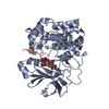
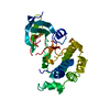
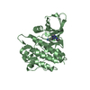
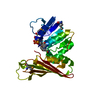
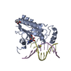
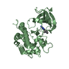
| タイトル | Molecular mechanism for the regulation of protein kinase B/Akt by hydrophobic motif phosphorylation |
|---|
 要素 要素 | RAC-BETA SERINE/THREONINE PROTEIN KINASE |
|---|
 キーワード キーワード | KINASE / TRANSFERASE / SERINE/THREONINE-PROTEIN KINASE / ATP-BINDING |
|---|
| 機能・相同性 |  機能・相同性情報 機能・相同性情報
retinal rod cell apoptotic process / PDE3B signalling / cellular response to high light intensity / Inhibition of TSC complex formation by PKB / positive regulation of cap-dependent translational initiation / AKT-mediated inactivation of FOXO1A / negative regulation of long-chain fatty acid import across plasma membrane / Negative regulation of the PI3K/AKT network / Activation of AKT2 / AKT phosphorylates targets in the nucleus ...retinal rod cell apoptotic process / PDE3B signalling / cellular response to high light intensity / Inhibition of TSC complex formation by PKB / positive regulation of cap-dependent translational initiation / AKT-mediated inactivation of FOXO1A / negative regulation of long-chain fatty acid import across plasma membrane / Negative regulation of the PI3K/AKT network / Activation of AKT2 / AKT phosphorylates targets in the nucleus / RUNX2 regulates genes involved in cell migration / positive regulation of fatty acid beta-oxidation / mammary gland epithelial cell differentiation / positive regulation of glucose metabolic process / RAB GEFs exchange GTP for GDP on RABs / peripheral nervous system myelin maintenance / glycogen biosynthetic process / AKT phosphorylates targets in the cytosol / Regulation of TP53 Activity through Association with Co-factors / positive regulation of cell motility / Co-inhibition by CTLA4 / Constitutive Signaling by AKT1 E17K in Cancer / fat cell differentiation / negative regulation of PERK-mediated unfolded protein response / Regulation of MITF-M-dependent genes involved in pigmentation / Regulation of localization of FOXO transcription factors / positive regulation of protein targeting to membrane / CD28 dependent PI3K/Akt signaling / Activation of BAD and translocation to mitochondria / Estrogen-dependent nuclear events downstream of ESR-membrane signaling / positive regulation of glycogen biosynthetic process / SARS-CoV-2 targets host intracellular signalling and regulatory pathways / Cyclin E associated events during G1/S transition / Cyclin A:Cdk2-associated events at S phase entry / Regulation of TP53 Activity through Acetylation / FLT3 Signaling / Downregulation of ERBB2:ERBB3 signaling / regulation of cell migration / molecular function activator activity / VEGFR2 mediated vascular permeability / protein localization to plasma membrane / Translocation of SLC2A4 (GLUT4) to the plasma membrane / positive regulation of D-glucose import / TP53 Regulates Metabolic Genes / Deactivation of the beta-catenin transactivating complex / protein modification process / ruffle membrane / Regulation of PTEN stability and activity / cellular response to insulin stimulus / glucose metabolic process / KEAP1-NFE2L2 pathway / G beta:gamma signalling through PI3Kgamma / Regulation of TP53 Degradation / insulin receptor signaling pathway / PIP3 activates AKT signaling / cell cortex / eukaryotic translation initiation factor 2alpha kinase activity / 3-phosphoinositide-dependent protein kinase activity / DNA-dependent protein kinase activity / ribosomal protein S6 kinase activity / histone H3S10 kinase activity / histone H2AXS139 kinase activity / histone H3S28 kinase activity / histone H4S1 kinase activity / histone H2BS14 kinase activity / histone H3T3 kinase activity / histone H2AS121 kinase activity / Rho-dependent protein serine/threonine kinase activity / histone H2BS36 kinase activity / histone H3S57 kinase activity / histone H2AT120 kinase activity / AMP-activated protein kinase activity / histone H2AS1 kinase activity / histone H3T6 kinase activity / early endosome / histone H3T11 kinase activity / histone H3T45 kinase activity / non-specific serine/threonine protein kinase / regulation of cell cycle / protein stabilization / intracellular signal transduction / positive regulation of cell migration / protein serine kinase activity / protein serine/threonine kinase activity / intracellular membrane-bounded organelle / signal transduction / nucleoplasm / ATP binding / metal ion binding / nucleus / plasma membrane / cytosol / cytoplasm類似検索 - 分子機能 Protein Kinase B beta, catalytic domain / Protein Kinase B, pleckstrin homology domain / Protein kinase, C-terminal / Protein kinase C terminal domain / Extension to Ser/Thr-type protein kinases / AGC-kinase, C-terminal / AGC-kinase C-terminal domain profile. / PH domain / PH domain profile. / Pleckstrin homology domain. ...Protein Kinase B beta, catalytic domain / Protein Kinase B, pleckstrin homology domain / Protein kinase, C-terminal / Protein kinase C terminal domain / Extension to Ser/Thr-type protein kinases / AGC-kinase, C-terminal / AGC-kinase C-terminal domain profile. / PH domain / PH domain profile. / Pleckstrin homology domain. / Pleckstrin homology domain / PH-like domain superfamily / Phosphorylase Kinase; domain 1 / Phosphorylase Kinase; domain 1 / Transferase(Phosphotransferase) domain 1 / Transferase(Phosphotransferase); domain 1 / Serine/threonine-protein kinase, active site / Serine/Threonine protein kinases active-site signature. / Protein kinase domain / Serine/Threonine protein kinases, catalytic domain / Protein kinase, ATP binding site / Protein kinases ATP-binding region signature. / Protein kinase domain profile. / Protein kinase domain / Protein kinase-like domain superfamily / 2-Layer Sandwich / Orthogonal Bundle / Mainly Alpha / Alpha Beta類似検索 - ドメイン・相同性 |
|---|
| 生物種 |  HOMO SAPIENS (ヒト) HOMO SAPIENS (ヒト) |
|---|
| 手法 |  X線回折 / シンクロトロン / 分子置換 / 解像度: 2.3 Å X線回折 / シンクロトロン / 分子置換 / 解像度: 2.3 Å |
|---|
 データ登録者 データ登録者 | Barford, D. / Yang, J. / Hemmings, B.A. |
|---|
 引用 引用 | ジャーナル: Mol.Cell / 年: 2002
タイトル: Molecular Mechanism for the Regulation of Protein Kinase B/Akt by Hydrophobic Motif Phosphorylation
著者: Yang, J. / Cron, P. / Thompson, V. / Good, V. / Hess, D. / Hemmings, B.A. / Barford, D. |
|---|
| 履歴 | | 登録 | 2002年5月23日 | 登録サイト: PDBE / 処理サイト: PDBE |
|---|
| 改定 1.0 | 2003年5月22日 | Provider: repository / タイプ: Initial release |
|---|
| 改定 1.1 | 2011年5月8日 | Group: Version format compliance |
|---|
| 改定 1.2 | 2011年7月13日 | Group: Version format compliance |
|---|
| 改定 1.3 | 2023年12月13日 | Group: Data collection / Database references ...Data collection / Database references / Other / Refinement description
カテゴリ: chem_comp_atom / chem_comp_bond ...chem_comp_atom / chem_comp_bond / database_2 / pdbx_database_status / pdbx_initial_refinement_model
Item: _database_2.pdbx_DOI / _database_2.pdbx_database_accession / _pdbx_database_status.status_code_sf |
|---|
|
|---|
 ムービー
ムービー コントローラー


 データを開く
 基本情報
基本情報 構造の表示
構造の表示 ダウンロードとリンク
 その他のダウンロード
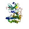


 PDBj
PDBj


 集合体
集合体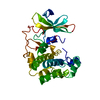

 SPODOPTERA FRUGIPERDA (ツマジロクサヨトウ)
 分子量: 18.015 Da / 分子数: 284 / 由来タイプ: 天然 / 式: H2O
 試料調製
試料調製 / ビームライン: ID14-4 / 波長: 0.9792
  解析
解析