 Movie
Movie Controller

[English] 日本語
 Yorodumi
- PDB-1gzc: High-Resolution crystal structure of Erythrina cristagalli lectin... -
+ Open data

- Basic information
| Entry | Database: PDB / ID: 1gzc | |||||||||
|---|---|---|---|---|---|---|---|---|---|---|
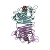
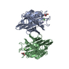
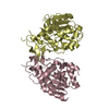
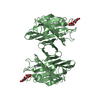
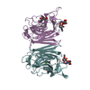
| Title | High-Resolution crystal structure of Erythrina cristagalli lectin in complex with lactose | |||||||||
 Components Components | ERYTHRINA CRISTA-GALLI LECTIN | |||||||||
 Keywords Keywords | SUGAR BINDING PROTEIN / LECTIN / CARBOHYDRATE / SACCHARIDE / PROTEIN-CARBOHYDRATE INTERACTIONS / LACTOSE / GLYCOPROTEIN | |||||||||
| Function / homology |  Function and homology information Function and homology information | |||||||||
| Biological species |  ERYTHRINA CRISTA-GALLI (cockspur coraltree) ERYTHRINA CRISTA-GALLI (cockspur coraltree) | |||||||||
| Method |  X-RAY DIFFRACTION / SYNCHROTRON / MOLECULAR REPLACEMENT / Resolution: 1.58 Å X-RAY DIFFRACTION / SYNCHROTRON / MOLECULAR REPLACEMENT / Resolution: 1.58 Å | |||||||||
 Authors Authors | Svensson, C. / Krengel, U. | |||||||||
 Citation Citation | Journal: J.Mol.Biol. / Year: 2002 Title: High-Resolution Crystal Structures of Erythrina Cristagalli Lectin in Complex with Lactose and 2'-Alpha-L-Fucosyllactose and Correlation with Thermodynamic Binding Data Authors: Svensson, C. / Teneberg, S. / Nilsson, C. / Kjellberg, A. / Schwarz, F. / Sharon, N. / Krengel, U. | |||||||||
| History |
| |||||||||
| Remark 700 | SHEET THE SHEET STRUCTURE OF THIS MOLECULE IS BIFURCATED. IN ORDER TO REPRESENT THIS FEATURE IN ... SHEET THE SHEET STRUCTURE OF THIS MOLECULE IS BIFURCATED. IN ORDER TO REPRESENT THIS FEATURE IN THE SHEET RECORDS BELOW, TWO SHEETS ARE DEFINED. |
- Structure visualization
| Structure viewer | Molecule: MolmilJmol/JSmol |
|---|
- Downloads & links
-Download
| PDBx/mmCIF format | 1gzc.cif.gz | 70.5 KB | Display | PDBx/mmCIF format |
|---|---|---|---|---|
| PDB format | pdb1gzc.ent.gz | 50.3 KB | Display | PDB format |
| PDBx/mmJSON format | 1gzc.json.gz | Tree view | PDBx/mmJSON format | |
| Others |  Other downloads Other downloads |
-Validation report
| Arichive directory | https://data.pdbj.org/pub/pdb/validation_reports/gz/1gzcftp://data.pdbj.org/pub/pdb/validation_reports/gz/1gzc | HTTPS FTP |
|---|
-Related structure data
| Related structure data | 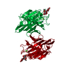 1gz9C 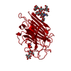 1ax1S C: citing same article ( S: Starting model for refinement |
|---|---|
| Similar structure data |


-Links
 PDBj
PDBj


- Assembly
| Deposited unit | 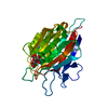
| ||||||||
|---|---|---|---|---|---|---|---|---|---|
| 1 | 
| ||||||||
| Unit cell |
|
-Components
| #1: Protein | Mass: 26249.189 Da / Num. of mol.: 1 / Source method: isolated from a natural source Source: (natural) ERYTHRINA CRISTA-GALLI (cockspur coraltree)References: UniProt: P83410*PLUS |
|---|---|
| #2: Polysaccharide | beta-D-galactopyranose-(1-4)-beta-D-glucopyranose / beta-lactose  Source method: isolated from a genetically manipulated source Details: oligosaccharide / References: beta-lactose |
| #3: Chemical | ChemComp-MN /   Mass: 54.938 Da / Num. of mol.: 1 / Source method: obtained synthetically / Formula: Mn Mass: 54.938 Da / Num. of mol.: 1 / Source method: obtained synthetically / Formula: Mn |
| #4: Chemical | ChemComp-CA /   Mass: 40.078 Da / Num. of mol.: 1 / Source method: obtained synthetically / Formula: Ca Mass: 40.078 Da / Num. of mol.: 1 / Source method: obtained synthetically / Formula: Ca |
| #5: Water | ChemComp-HOH /  Mass: 18.015 Da / Num. of mol.: 266 / Source method: isolated from a natural source / Formula: H2O Mass: 18.015 Da / Num. of mol.: 266 / Source method: isolated from a natural source / Formula: H2O |
| Sequence details | THE SEQUENCE OF THE CRYSTALLIZED PROTEIN WAS CONFIRMED BY MASS SPECTROMETRY, BUT THERE ARE ...THE SEQUENCE OF THE CRYSTALLIZ |
-Experimental details
-Experiment
| Experiment | Method: X-RAY DIFFRACTION / Number of used crystals: 1 |
|---|
- Sample preparation
| Crystal | Density Matthews: 4 Å3/Da / Density % sol: 69 % Description: THE DATA SET INCLUDED REFLECTIONS TO 1.45 A RESOLUTION, BUT THE DATA WERE CUT TO 1.58 A RESOLUTION IN THE FINAL REFINEMENT CYCLE DUE TO CONSIDERABLE INCREASE IN R-FACTORS BEYOND THAT RESOLUTION. | |||||||||||||||||||||||||||||||||||||||||||||||||
|---|---|---|---|---|---|---|---|---|---|---|---|---|---|---|---|---|---|---|---|---|---|---|---|---|---|---|---|---|---|---|---|---|---|---|---|---|---|---|---|---|---|---|---|---|---|---|---|---|---|---|
| Crystal grow | pH: 7.5 Details: 2M AMMONIUM SULFATE, 0.1M TRIS PH 7.5, 10% GLYCEROL | |||||||||||||||||||||||||||||||||||||||||||||||||
| Crystal grow | *PLUS pH: 8 / Method: vapor diffusion, hanging drop / Details: used macroseeding | |||||||||||||||||||||||||||||||||||||||||||||||||
| Components of the solutions | *PLUS
|
-Data collection
| Diffraction | Mean temperature: 100 K |
|---|---|
| Diffraction source | Source: SYNCHROTRON / Site: MAX II  / Beamline: I711 / Wavelength: 1.03 / Beamline: I711 / Wavelength: 1.03 |
| Detector | Type: MARRESEARCH / Detector: IMAGE PLATE / Date: Oct 15, 2000 |
| Radiation | Protocol: SINGLE WAVELENGTH / Monochromatic (M) / Laue (L): M / Scattering type: x-ray |
| Radiation wavelength | Wavelength: 1.03 Å / Relative weight: 1 |
| Reflection | Resolution: 1.58→49.8 Å / Num. obs: 58614 / % possible obs: 99.5 % / Redundancy: 9.4 % / Biso Wilson estimate: 22.6 Å2 / Rmerge(I) obs: 0.053 / Net I/σ(I): 20.9 |
| Reflection shell | Resolution: 1.58→1.64 Å / Redundancy: 6.4 % / Rmerge(I) obs: 0.608 / Mean I/σ(I) obs: 2.3 / % possible all: 99.4 |
| Reflection | *PLUS Highest resolution: 1.6 Å / Num. measured all: 551905 |
| Reflection shell | *PLUS % possible obs: 99.4 % / Num. unique obs: 6065 / Num. measured obs: 38515 |
- Processing
| Software |
| ||||||||||||||||||||||||||||||||||||||||||||||||||||||||||||||||||||||||||||||||
|---|---|---|---|---|---|---|---|---|---|---|---|---|---|---|---|---|---|---|---|---|---|---|---|---|---|---|---|---|---|---|---|---|---|---|---|---|---|---|---|---|---|---|---|---|---|---|---|---|---|---|---|---|---|---|---|---|---|---|---|---|---|---|---|---|---|---|---|---|---|---|---|---|---|---|---|---|---|---|---|---|---|
| Refinement | Method to determine structure: MOLECULAR REPLACEMENT Starting model: 1AX1 Resolution: 1.58→49.9 Å / Isotropic thermal model: RESTRAINED / Cross valid method: THROUGHOUT / σ(F): 0 Details: ALTERNATIVE CONFORMATIONS REFINED FOR RESIDUES 9, 10, 12, 95, 159, 173, 180, 234 LACTOSE ARE DEPOSITED: LAT_XPLOR_TOP.TXT LAT_XPLOR_PAR.TXT. INITIAL STAGES OF REFINEMENT WITH SIMULATED ANNEALING.
| ||||||||||||||||||||||||||||||||||||||||||||||||||||||||||||||||||||||||||||||||
| Solvent computation | Bsol: 51.5279 Å2 / ksol: 0.385527 e/Å3 | ||||||||||||||||||||||||||||||||||||||||||||||||||||||||||||||||||||||||||||||||
| Displacement parameters | Biso mean: 27.4 Å2
| ||||||||||||||||||||||||||||||||||||||||||||||||||||||||||||||||||||||||||||||||
| Refinement step | Cycle: LAST / Resolution: 1.58→49.9 Å
| ||||||||||||||||||||||||||||||||||||||||||||||||||||||||||||||||||||||||||||||||
| Refine LS restraints |
| ||||||||||||||||||||||||||||||||||||||||||||||||||||||||||||||||||||||||||||||||
| LS refinement shell | Resolution: 1.58→1.59 Å / Total num. of bins used: 50
| ||||||||||||||||||||||||||||||||||||||||||||||||||||||||||||||||||||||||||||||||
| Xplor file |
| ||||||||||||||||||||||||||||||||||||||||||||||||||||||||||||||||||||||||||||||||
| Refinement | *PLUS Highest resolution: 1.6 Å | ||||||||||||||||||||||||||||||||||||||||||||||||||||||||||||||||||||||||||||||||
| Solvent computation | *PLUS | ||||||||||||||||||||||||||||||||||||||||||||||||||||||||||||||||||||||||||||||||
| Displacement parameters | *PLUS | ||||||||||||||||||||||||||||||||||||||||||||||||||||||||||||||||||||||||||||||||
| LS refinement shell | *PLUS Lowest resolution: 1.64 Å |
