 Movie
Movie Controller


+ Open data

- Basic information
| Entry | Database: PDB / ID: 1gxr | ||||||
|---|---|---|---|---|---|---|---|
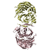
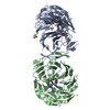
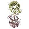
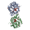
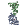
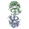
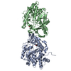
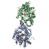
| Title | WD40 Region of Human Groucho/TLE1 | ||||||
 Components Components | TRANSDUCIN-LIKE ENHANCER PROTEIN 1 | ||||||
 Keywords Keywords | TRANSCRIPTION / TRANSCRIPTIONAL CO-REPRESSOR / WD40 / TRANSCRIPTION REPRESSOR / WD REPEAT | ||||||
| Function / homology |  Function and homology information Function and homology informationRepression of WNT target genes / beta-catenin-TCF complex / negative regulation of Wnt signaling pathway / negative regulation of anoikis / animal organ morphogenesis / negative regulation of canonical NF-kappaB signal transduction / Deactivation of the beta-catenin transactivating complex / negative regulation of canonical Wnt signaling pathway / Formation of the beta-catenin:TCF transactivating complex / Negative Regulation of CDH1 Gene Transcription ...Repression of WNT target genes / beta-catenin-TCF complex / negative regulation of Wnt signaling pathway / negative regulation of anoikis / animal organ morphogenesis / negative regulation of canonical NF-kappaB signal transduction / Deactivation of the beta-catenin transactivating complex / negative regulation of canonical Wnt signaling pathway / Formation of the beta-catenin:TCF transactivating complex / Negative Regulation of CDH1 Gene Transcription / NOTCH1 Intracellular Domain Regulates Transcription / Wnt signaling pathway / transcription corepressor activity / transcription regulator complex / DNA-binding transcription factor binding / negative regulation of DNA-templated transcription / positive regulation of gene expression / signal transduction / nucleoplasm / identical protein binding / nucleus / cytosol Similarity search - Function | ||||||
| Biological species |  HOMO SAPIENS (human) HOMO SAPIENS (human) | ||||||
| Method |  X-RAY DIFFRACTION / SYNCHROTRON / MOLECULAR REPLACEMENT / Resolution: 1.65 Å X-RAY DIFFRACTION / SYNCHROTRON / MOLECULAR REPLACEMENT / Resolution: 1.65 Å | ||||||
 Authors Authors | Pearl, L.H. / Roe, S.M. / Pickles, L.M. | ||||||
 Citation Citation | Journal: Structure / Year: 2002 Title: Crystal Structure of the C-Terminal Wd40 Repeat Domain of the Human Groucho/Tle1 Transcriptional Corepressor Authors: Pickles, L.M. / Roe, S.M. / Hemingway, E.J. / Stifani, S. / Pearl, L.H. | ||||||
| History |
|
- Structure visualization
| Structure viewer | Molecule: MolmilJmol/JSmol |
|---|
- Downloads & links
-Download
| PDBx/mmCIF format | 1gxr.cif.gz | 150.2 KB | Display | PDBx/mmCIF format |
|---|---|---|---|---|
| PDB format | pdb1gxr.ent.gz | 116 KB | Display | PDB format |
| PDBx/mmJSON format | 1gxr.json.gz | Tree view | PDBx/mmJSON format | |
| Others |  Other downloads Other downloads |
-Validation report
| Arichive directory | https://data.pdbj.org/pub/pdb/validation_reports/gx/1gxrftp://data.pdbj.org/pub/pdb/validation_reports/gx/1gxr | HTTPS FTP |
|---|
-Related structure data
| Related structure data |  1erjS S: Starting model for refinement |
|---|---|
| Similar structure data |


-Links
 PDBj
PDBj- Assembly
| Deposited unit | 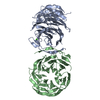
| ||||||||
|---|---|---|---|---|---|---|---|---|---|
| 1 |
| ||||||||
| Unit cell |
|
-Components
| #1: Protein | Mass: 36853.469 Da / Num. of mol.: 2 Fragment: RESIDUES 443-770 (END OF SP-REGION 443-473 AND WD40 REPEAT DOMAIN 474-770) Source method: isolated from a genetically manipulated source Details: THIS CONSTRUCT CONTAINS ALL THE SEVEN WD REPEATS / Source: (gene. exp.) HOMO SAPIENS (human) / Plasmid: PFASTBAC / Production host:   SPODOPTERA FRUGIPERDA (fall armyworm) / Strain (production host): SF9 / References: UniProt: Q04724 SPODOPTERA FRUGIPERDA (fall armyworm) / Strain (production host): SF9 / References: UniProt: Q04724#2: Chemical | ChemComp-CA / |   Mass: 40.078 Da / Num. of mol.: 1 / Source method: obtained synthetically / Formula: Ca Mass: 40.078 Da / Num. of mol.: 1 / Source method: obtained synthetically / Formula: Ca#3: Water | ChemComp-HOH / |  Mass: 18.015 Da / Num. of mol.: 564 / Source method: isolated from a natural source / Formula: H2O Mass: 18.015 Da / Num. of mol.: 564 / Source method: isolated from a natural source / Formula: H2OCompound details | CHAIN A, B RESIDUES 475-511 WD REPEAT 1 CHAIN A, B RESIDUES 522-558 WD REPEAT 2 CHAIN A, B RESIDUES ...CHAIN A, B RESIDUES 475-511 WD REPEAT 1 CHAIN A, B RESIDUES 522-558 WD REPEAT 2 CHAIN A, B RESIDUES 565-602 WD REPEAT 3 CHAIN A, B RESIDUES 607-644 WD REPEAT 4 CHAIN A, B RESIDUES 649-685 WD REPEAT 5 CHAIN A, B RESIDUES 791-726 WD REPEAT 6 CHAIN A, B RESIDUES 732-768 WD REPEAT 7 | Sequence details | THE CONFLICT IS CONFIRMED BY RESEQUENCING THE GENE AND THE RESIDUES 464 AND 465 ARE CONFIRMED TO BE ...THE CONFLICT IS CONFIRMED BY RESEQUENCI | |
|---|
-Experimental details
-Experiment
| Experiment | Method: X-RAY DIFFRACTION / Number of used crystals: 1 |
|---|
- Sample preparation
| Crystal | Density Matthews: 2.4 Å3/Da / Density % sol: 48 % | |||||||||||||||||||||||||
|---|---|---|---|---|---|---|---|---|---|---|---|---|---|---|---|---|---|---|---|---|---|---|---|---|---|---|
| Crystal grow | Temperature: 287 K / Method: microbatch / pH: 7 Details: CRYSTALS WERE GROWN BY MICROBATCH METHODS AT 14C. PROTEIN AT 6MG/ML, WAS MIXED WITH AN EQUAL VOLUME O PRECIPITANT (22% PEG8000, 100MM, NACACODYLATE, 100MM CAACETATE)., pH 7.00 | |||||||||||||||||||||||||
| Crystal grow | *PLUS Temperature: 14 ℃ / Method: microdialysis | |||||||||||||||||||||||||
| Components of the solutions | *PLUS
|
-Data collection
| Diffraction | Mean temperature: 100 K |
|---|---|
| Diffraction source | Source: SYNCHROTRON / Site: ESRF  / Beamline: ID14-2 / Wavelength: 0.933 / Beamline: ID14-2 / Wavelength: 0.933 |
| Detector | Type: ADSC CCD / Detector: CCD / Date: Jan 15, 2002 |
| Radiation | Protocol: SINGLE WAVELENGTH / Monochromatic (M) / Laue (L): M / Scattering type: x-ray |
| Radiation wavelength | Wavelength: 0.933 Å / Relative weight: 1 |
| Reflection | Resolution: 1.6→40 Å / Num. obs: 92007 / % possible obs: 100 % / Redundancy: 5.2 % / Biso Wilson estimate: 24 Å2 / Rmerge(I) obs: 0.064 / Net I/σ(I): 5.4 |
| Reflection shell | Resolution: 1.6→1.67 Å / Redundancy: 3.7 % / Rmerge(I) obs: 0.273 / Mean I/σ(I) obs: 2.6 / % possible all: 100 |
| Reflection | *PLUS Highest resolution: 1.65 Å / Lowest resolution: 50 Å / Num. obs: 83990 / % possible obs: 100 % |
| Reflection shell | *PLUS Highest resolution: 1.6 Å / Lowest resolution: 1.7 Å / % possible obs: 100 % |
- Processing
| Software |
| ||||||||||||||||||||||||||||||||||||||||||||||||||||||||||||||||||||||||||||||||
|---|---|---|---|---|---|---|---|---|---|---|---|---|---|---|---|---|---|---|---|---|---|---|---|---|---|---|---|---|---|---|---|---|---|---|---|---|---|---|---|---|---|---|---|---|---|---|---|---|---|---|---|---|---|---|---|---|---|---|---|---|---|---|---|---|---|---|---|---|---|---|---|---|---|---|---|---|---|---|---|---|---|
| Refinement | Method to determine structure: MOLECULAR REPLACEMENT Starting model: PDB ENTRY 1ERJ Resolution: 1.65→40.45 Å / Rfactor Rfree error: 0.004 / Data cutoff high absF: 1439675.18 / Isotropic thermal model: RESTRAINED / Cross valid method: THROUGHOUT / σ(F): 0
| ||||||||||||||||||||||||||||||||||||||||||||||||||||||||||||||||||||||||||||||||
| Solvent computation | Solvent model: FLAT MODEL / Bsol: 44.0585 Å2 / ksol: 0.348345 e/Å3 | ||||||||||||||||||||||||||||||||||||||||||||||||||||||||||||||||||||||||||||||||
| Displacement parameters | Biso mean: 32.2 Å2
| ||||||||||||||||||||||||||||||||||||||||||||||||||||||||||||||||||||||||||||||||
| Refine analyze |
| ||||||||||||||||||||||||||||||||||||||||||||||||||||||||||||||||||||||||||||||||
| Refinement step | Cycle: LAST / Resolution: 1.65→40.45 Å
| ||||||||||||||||||||||||||||||||||||||||||||||||||||||||||||||||||||||||||||||||
| Refine LS restraints |
| ||||||||||||||||||||||||||||||||||||||||||||||||||||||||||||||||||||||||||||||||
| LS refinement shell | Resolution: 1.65→1.75 Å / Rfactor Rfree error: 0.012 / Total num. of bins used: 6
| ||||||||||||||||||||||||||||||||||||||||||||||||||||||||||||||||||||||||||||||||
| Xplor file |
| ||||||||||||||||||||||||||||||||||||||||||||||||||||||||||||||||||||||||||||||||
| Refinement | *PLUS % reflection Rfree: 5 % / Rfactor obs: 0.23 / Rfactor Rwork: 0.23 | ||||||||||||||||||||||||||||||||||||||||||||||||||||||||||||||||||||||||||||||||
| Solvent computation | *PLUS | ||||||||||||||||||||||||||||||||||||||||||||||||||||||||||||||||||||||||||||||||
| Displacement parameters | *PLUS | ||||||||||||||||||||||||||||||||||||||||||||||||||||||||||||||||||||||||||||||||
| Refine LS restraints | *PLUS
| ||||||||||||||||||||||||||||||||||||||||||||||||||||||||||||||||||||||||||||||||
| LS refinement shell | *PLUS Rfactor obs: 0.268 |
