 Movie
Movie Controller

[English] 日本語
 Yorodumi
- PDB-1gxk: SMC hinge domain from T. maritima w/o coiled coil, P212121 crysta... -
+ Open data

- Basic information
| Entry | Database: PDB / ID: 1gxk | ||||||
|---|---|---|---|---|---|---|---|
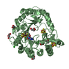
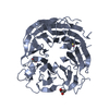
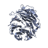
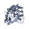
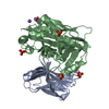
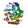
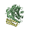
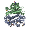
| Title | SMC hinge domain from T. maritima w/o coiled coil, P212121 crystal form | ||||||
 Components Components | CHROMOSOME SEGREGATION SMC PROTEIN | ||||||
 Keywords Keywords | CHROMOSOME SEGREGATION / SMC DIMERISATION DOMAIN / ANTI PARALLEL COILED COIL / SMC PROTEINS / COMPLETE PROTEOME. | ||||||
| Function / homology |  Function and homology information Function and homology informationchromosome condensation / sister chromatid cohesion / chromosome segregation / chromosome / DNA replication / ATP hydrolysis activity / DNA binding / ATP binding / identical protein binding / cytoplasm Similarity search - Function | ||||||
| Biological species |   THERMOTOGA MARITIMA (bacteria) THERMOTOGA MARITIMA (bacteria) | ||||||
| Method |  X-RAY DIFFRACTION / SYNCHROTRON / MOLECULAR REPLACEMENT / Resolution: 3 Å X-RAY DIFFRACTION / SYNCHROTRON / MOLECULAR REPLACEMENT / Resolution: 3 Å | ||||||
 Authors Authors | Lowe, J. / Haering, C. / Nasmyth, K. | ||||||
 Citation Citation | Journal: Mol.Cell / Year: 2002 Title: Molecular Architecture of Smc Proteins and the Yeast Cohesin Complex Authors: Haering, C. / Lowe, J. / Hochwagen, A. / Nasmyth, K. | ||||||
| History |
|
- Structure visualization
| Structure viewer | Molecule: MolmilJmol/JSmol |
|---|
- Downloads & links
-Download
| PDBx/mmCIF format | 1gxk.cif.gz | 130.4 KB | Display | PDBx/mmCIF format |
|---|---|---|---|---|
| PDB format | pdb1gxk.ent.gz | 103.3 KB | Display | PDB format |
| PDBx/mmJSON format | 1gxk.json.gz | Tree view | PDBx/mmJSON format | |
| Others |  Other downloads Other downloads |
-Validation report
| Arichive directory | https://data.pdbj.org/pub/pdb/validation_reports/gx/1gxkftp://data.pdbj.org/pub/pdb/validation_reports/gx/1gxk | HTTPS FTP |
|---|
-Related structure data
| Related structure data |  1gxjSC  1gxlC S: Starting model for refinement C: citing same article ( |
|---|---|
| Similar structure data |


-Links
 PDBj
PDBj- Assembly
| Deposited unit | 
| ||||||||
|---|---|---|---|---|---|---|---|---|---|
| 1 | 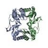
| ||||||||
| 2 | 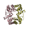
| ||||||||
| Unit cell |
|
-Components
| #1: Protein | Mass: 20844.791 Da / Num. of mol.: 4 / Fragment: HINGE DOMAIN RESIDUES 485-670 Source method: isolated from a genetically manipulated source Details: HINGE DOMAIN FROM T. MARITIMA, RESIDUES 485-670, ORTHORHOMBIC FORM Source: (gene. exp.) THERMOTOGA MARITIMA (bacteria) / Strain: DSMZ 3109 / Production host: |
|---|
-Experimental details
-Experiment
| Experiment | Method: X-RAY DIFFRACTION |
|---|
- Sample preparation
| Crystal | Density Matthews: 2.47 Å3/Da / Density % sol: 45 % | ||||||||||||||||||||||||
|---|---|---|---|---|---|---|---|---|---|---|---|---|---|---|---|---|---|---|---|---|---|---|---|---|---|
| Crystal grow | pH: 9.2 / Details: pH 9.20 | ||||||||||||||||||||||||
| Crystal grow | *PLUS Temperature: 19 ℃ / Method: vapor diffusion, sitting drop / pH: 6.9 | ||||||||||||||||||||||||
| Components of the solutions | *PLUS
|
-Data collection
| Diffraction | Mean temperature: 100 K |
|---|---|
| Diffraction source | Source: SYNCHROTRON / Site: ESRF  / Beamline: BM14 / Wavelength: 0.93 / Beamline: BM14 / Wavelength: 0.93 |
| Radiation | Protocol: SINGLE WAVELENGTH / Monochromatic (M) / Laue (L): M / Scattering type: x-ray |
| Radiation wavelength | Wavelength: 0.93 Å / Relative weight: 1 |
| Reflection | Resolution: 3→30 Å / Num. obs: 13536 / % possible obs: 92.7 % / Redundancy: 2.8 % / Biso Wilson estimate: 44 Å2 / Rmerge(I) obs: 0.093 / Net I/σ(I): 10.6 |
| Reflection shell | Resolution: 3→3.16 Å / Redundancy: 2.8 % / Rmerge(I) obs: 0.235 / Mean I/σ(I) obs: 3.1 / % possible all: 92.6 |
- Processing
| Software |
| ||||||||||||||||||||||||||||||||||||||||||||||||||||||||||||
|---|---|---|---|---|---|---|---|---|---|---|---|---|---|---|---|---|---|---|---|---|---|---|---|---|---|---|---|---|---|---|---|---|---|---|---|---|---|---|---|---|---|---|---|---|---|---|---|---|---|---|---|---|---|---|---|---|---|---|---|---|---|
| Refinement | Method to determine structure: MOLECULAR REPLACEMENT Starting model: P21 CRYSTAL FORM, PDB ID 1GXJ Resolution: 3→30 Å / Cross valid method: THROUGHOUT / σ(F): 0 Details: DATA IS TWINNED. TWIN OPERATOR K, H, - TWIN FRACTION 0.158
| ||||||||||||||||||||||||||||||||||||||||||||||||||||||||||||
| Displacement parameters | Biso mean: 49.68 Å2 | ||||||||||||||||||||||||||||||||||||||||||||||||||||||||||||
| Refinement step | Cycle: LAST / Resolution: 3→30 Å
| ||||||||||||||||||||||||||||||||||||||||||||||||||||||||||||
| Refine LS restraints |
| ||||||||||||||||||||||||||||||||||||||||||||||||||||||||||||
| Refine LS restraints NCS | NCS model details: RESTRAINT / Rms dev position: 0.1084 Å / Weight position: 300 | ||||||||||||||||||||||||||||||||||||||||||||||||||||||||||||
| Refinement | *PLUS % reflection Rfree: 5 % / Rfactor obs: 0.253 / Rfactor Rfree: 0.298 / Rfactor Rwork: 0.253 | ||||||||||||||||||||||||||||||||||||||||||||||||||||||||||||
| Solvent computation | *PLUS | ||||||||||||||||||||||||||||||||||||||||||||||||||||||||||||
| Displacement parameters | *PLUS |
