 Movie
Movie Controller


+ Open data

- Basic information
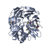
| Entry | Database: PDB / ID: 6n01 | ||||||
|---|---|---|---|---|---|---|---|
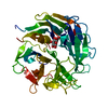
| Title | Structure of apo AztD from Citrobacter koseri | ||||||
 Components Components | AztD Protein | ||||||
 Keywords Keywords | CHAPERONE / periplasm / beta propeller / zinc / metallochaperone | ||||||
| Function / homology | : / Nitrous oxide reductase, N-terminal / periplasmic space / WD40/YVTN repeat-like-containing domain superfamily / metal ion binding / TRIETHYLENE GLYCOL / Zinc chaperone AztD Function and homology information Function and homology information | ||||||
| Biological species |  Citrobacter koseri (bacteria) Citrobacter koseri (bacteria) | ||||||
| Method |  X-RAY DIFFRACTION / SYNCHROTRON / MOLECULAR REPLACEMENT / Resolution: 1.98 Å X-RAY DIFFRACTION / SYNCHROTRON / MOLECULAR REPLACEMENT / Resolution: 1.98 Å | ||||||
 Authors Authors | Yukl, E.T. / Neupane, D.P. | ||||||
| Funding support |  United States, 1items United States, 1items
| ||||||
 Citation Citation | Journal: Commun Biol / Year: 2019 Title: Crystal structures of AztD provide mechanistic insights into direct zinc transfer between proteins. Authors: Neupane, D.P. / Fullam, S.H. / Chacon, K.N. / Yukl, E.T. | ||||||
| History |
|
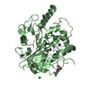
- Structure visualization
| Structure viewer | Molecule: MolmilJmol/JSmol |
|---|
- Downloads & links
-Download
| PDBx/mmCIF format | 6n01.cif.gz | 523.4 KB | Display | PDBx/mmCIF format |
|---|---|---|---|---|
| PDB format | pdb6n01.ent.gz | 433.8 KB | Display | PDB format |
| PDBx/mmJSON format | 6n01.json.gz | Tree view | PDBx/mmJSON format | |
| Others |  Other downloads Other downloads |
-Validation report
| Arichive directory | https://data.pdbj.org/pub/pdb/validation_reports/n0/6n01ftp://data.pdbj.org/pub/pdb/validation_reports/n0/6n01 | HTTPS FTP |
|---|
-Related structure data
| Related structure data | 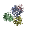 6ck1C 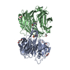 6cmkSC S: Starting model for refinement C: citing same article ( |
|---|---|
| Similar structure data |


-Links
 PDBj
PDBj
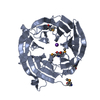
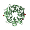
- Assembly
| Deposited unit | 
| ||||||||
|---|---|---|---|---|---|---|---|---|---|
| 1 | 
| ||||||||
| 2 | 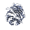
| ||||||||
| 3 | 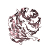
| ||||||||
| 4 | 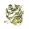
| ||||||||
| Unit cell |
|
-Components
| #1: Protein | Mass: 45467.371 Da / Num. of mol.: 4 Source method: isolated from a genetically manipulated source Source: (gene. exp.) Citrobacter koseri (strain ATCC BAA-895 / CDC 4225-83 / SGSC4696) (bacteria)Strain: ATCC BAA-895 / CDC 4225-83 / SGSC4696 / Gene: CKO_00948 / Production host: #2: Chemical |   Mass: 150.173 Da / Num. of mol.: 2 / Source method: obtained synthetically / Formula: C6H14O4 Mass: 150.173 Da / Num. of mol.: 2 / Source method: obtained synthetically / Formula: C6H14O4#3: Chemical |   Mass: 62.068 Da / Num. of mol.: 2 / Source method: obtained synthetically / Formula: C2H6O2 Mass: 62.068 Da / Num. of mol.: 2 / Source method: obtained synthetically / Formula: C2H6O2#4: Chemical | ChemComp-MES / |   Mass: 195.237 Da / Num. of mol.: 1 / Source method: obtained synthetically / Formula: C6H13NO4S / Comment: pH buffer*YM Mass: 195.237 Da / Num. of mol.: 1 / Source method: obtained synthetically / Formula: C6H13NO4S / Comment: pH buffer*YM#5: Water | ChemComp-HOH / |  Mass: 18.015 Da / Num. of mol.: 501 / Source method: isolated from a natural source / Formula: H2O Mass: 18.015 Da / Num. of mol.: 501 / Source method: isolated from a natural source / Formula: H2OHas protein modification | Y | |
|---|
-Experimental details
-Experiment
| Experiment | Method: X-RAY DIFFRACTION / Number of used crystals: 1 |
|---|
- Sample preparation
| Crystal | Density Matthews: 2.25 Å3/Da / Density % sol: 45.45 % |
|---|---|
| Crystal grow | Temperature: 292 K / Method: batch mode / pH: 6 Details: 10 mg/mL protein was combined in a 1:1 ratio with 24% PEG 4000, 0.1 M sodium nitrate, 0.1 M MES pH 6.0 |
-Data collection
| Diffraction | Mean temperature: 100 K / Serial crystal experiment: N |
|---|---|
| Diffraction source | Source: SYNCHROTRON / Site: ALS / Beamline: 5.0.2 / Wavelength: 1 Å |
| Detector | Type: DECTRIS PILATUS3 S 6M / Detector: PIXEL / Date: Jun 30, 2018 |
| Radiation | Monochromator: Double-crystal, Si(111) / Protocol: SINGLE WAVELENGTH / Monochromatic (M) / Laue (L): M / Scattering type: x-ray |
| Radiation wavelength | Wavelength: 1 Å / Relative weight: 1 |
| Reflection | Resolution: 1.98→48.43 Å / Num. obs: 110365 / % possible obs: 98.6 % / Redundancy: 3.4 % / CC1/2: 0.997 / Rmerge(I) obs: 0.072 / Rpim(I) all: 0.044 / Rrim(I) all: 0.085 / Χ2: 0.9 / Net I/σ(I): 9.7 |
| Reflection shell | Resolution: 1.98→2.01 Å / Redundancy: 3.1 % / Rmerge(I) obs: 0.524 / Mean I/σ(I) obs: 2.1 / Num. unique obs: 16782 / CC1/2: 0.758 / Rpim(I) all: 0.326 / Rrim(I) all: 0.621 / Χ2: 0.69 / % possible all: 96.4 |
- Processing
| Software |
| |||||||||||||||||||||||||||||||||||||||||||||||||||||||||||||||||||||||||||||||||||||||||||||||||||||||||
|---|---|---|---|---|---|---|---|---|---|---|---|---|---|---|---|---|---|---|---|---|---|---|---|---|---|---|---|---|---|---|---|---|---|---|---|---|---|---|---|---|---|---|---|---|---|---|---|---|---|---|---|---|---|---|---|---|---|---|---|---|---|---|---|---|---|---|---|---|---|---|---|---|---|---|---|---|---|---|---|---|---|---|---|---|---|---|---|---|---|---|---|---|---|---|---|---|---|---|---|---|---|---|---|---|---|---|
| Refinement | Method to determine structure: MOLECULAR REPLACEMENT Starting model: 6cmk Resolution: 1.98→48.431 Å / SU ML: 0.24 / Cross valid method: FREE R-VALUE / σ(F): 1.34 / Phase error: 26.85 / Stereochemistry target values: ML
| |||||||||||||||||||||||||||||||||||||||||||||||||||||||||||||||||||||||||||||||||||||||||||||||||||||||||
| Solvent computation | Shrinkage radii: 0.9 Å / VDW probe radii: 1.11 Å / Solvent model: FLAT BULK SOLVENT MODEL | |||||||||||||||||||||||||||||||||||||||||||||||||||||||||||||||||||||||||||||||||||||||||||||||||||||||||
| Refinement step | Cycle: LAST / Resolution: 1.98→48.431 Å
| |||||||||||||||||||||||||||||||||||||||||||||||||||||||||||||||||||||||||||||||||||||||||||||||||||||||||
| Refine LS restraints |
| |||||||||||||||||||||||||||||||||||||||||||||||||||||||||||||||||||||||||||||||||||||||||||||||||||||||||
| LS refinement shell |
|
