 Movie
Movie Controller

[English] 日本語
 Yorodumi
- PDB-1gtk: Time-resolved and static-ensemble structural chemistry of hydroxy... -
+ Open data

- Basic information
| Entry | Database: PDB / ID: 1gtk | ||||||
|---|---|---|---|---|---|---|---|
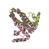
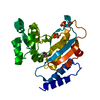
| Title | Time-resolved and static-ensemble structural chemistry of hydroxymethylbilane synthase | ||||||
 Components Components | PORPHOBILINOGEN DEAMINASE | ||||||
 Keywords Keywords | TRANSFERASE / BIOSYNTHESIS OF LINEAR TETRAPYRROLE / ALL ALPHA/BETA | ||||||
| Function / homology |  Function and homology information Function and homology informationtetrapyrrole biosynthetic process / hydroxymethylbilane synthase / hydroxymethylbilane synthase activity / : / heme biosynthetic process / cytoplasm / cytosol Similarity search - Function | ||||||
| Biological species |  | ||||||
| Method |  X-RAY DIFFRACTION / SYNCHROTRON / MOLECULAR REPLACEMENT / Resolution: 1.66 Å X-RAY DIFFRACTION / SYNCHROTRON / MOLECULAR REPLACEMENT / Resolution: 1.66 Å | ||||||
 Authors Authors | Helliwell, J.R. / Nieh, Y.P. / Raftery, J. / Cassetta, A. / Habash, J. / Carr, P.D. / Ursby, T. / Wulff, M. / Thompson, A.W. / Niemann, A.C. / Haedener, A. | ||||||
 Citation Citation | Journal: Faraday Discuss. / Year: 2003 Title: Time-Resolved and Static-Ensemble Structural Chemistry of Hydroxymethylbilane Synthase Authors: Helliwell, J.R. / Nieh, Y.P. / Habash, J. / Faulder, P.F. / Raftery, J. / Cianci, M. / Wulff, M. / Hadener, A. #1: Journal: J.Chem.Soc.,Faraday Trans. / Year: 1998Title: Time-Resolved Structures of Hydroxymethylbilane Synthase (Lys59Gln Mutant) as It is Loaded with Substrate in the Crystal Determined by Laue Diffraction Authors: Helliwell, J.R. / Nieh, Y.P. / Raftery, J. / Cassetta, A. / Habash, J. / Carr, P.D. / Ursby, T. / Wulff, M. / Thompson, A.W. / Niemann, A.C. / Hadener, A. | ||||||
| History |
|
- Structure visualization
| Structure viewer | Molecule: MolmilJmol/JSmol |
|---|
- Downloads & links
-Download
| PDBx/mmCIF format | 1gtk.cif.gz | 80 KB | Display | PDBx/mmCIF format |
|---|---|---|---|---|
| PDB format | pdb1gtk.ent.gz | 57.9 KB | Display | PDB format |
| PDBx/mmJSON format | 1gtk.json.gz | Tree view | PDBx/mmJSON format | |
| Others |  Other downloads Other downloads |
-Validation report
| Arichive directory | https://data.pdbj.org/pub/pdb/validation_reports/gt/1gtkftp://data.pdbj.org/pub/pdb/validation_reports/gt/1gtk | HTTPS FTP |
|---|
-Related structure data
| Related structure data |  2ypnS S: Starting model for refinement |
|---|---|
| Similar structure data |


-Links
 PDBj
PDBj- Assembly
| Deposited unit | 
| ||||||||
|---|---|---|---|---|---|---|---|---|---|
| 1 |
| ||||||||
| Unit cell |
| ||||||||
| Components on special symmetry positions |
|
-Components
| #1: Protein | Mass: 33891.805 Da / Num. of mol.: 1 / Fragment: THREE DOMAINS Source method: isolated from a genetically manipulated source Details: CONTAINS A DIPYRROMETHANE COFACTOR LINKED TO CYSTEINE 242 Source: (gene. exp.) |
|---|---|
| #2: Chemical | ChemComp-DPM /   Mass: 420.413 Da / Num. of mol.: 1 / Source method: obtained synthetically / Formula: C20H24N2O8 Mass: 420.413 Da / Num. of mol.: 1 / Source method: obtained synthetically / Formula: C20H24N2O8 |
| #3: Water | ChemComp-HOH /  Mass: 18.015 Da / Num. of mol.: 320 / Source method: isolated from a natural source / Formula: H2O Mass: 18.015 Da / Num. of mol.: 320 / Source method: isolated from a natural source / Formula: H2O |
-Experimental details
-Experiment
| Experiment | Method: X-RAY DIFFRACTION / Number of used crystals: 1 |
|---|
- Sample preparation
| Crystal | Density Matthews: 2.45 Å3/Da / Density % sol: 50 % Description: THE STRUCTURE WAS ALREADY DETERMINED BUT REFINED HERE ONLY |
|---|---|
| Crystal grow | Method: vapor diffusion, sitting drop / pH: 5.3 Details: PROTEIN WAS CRYSTALLISED AT PH 5.3 IN SITTING DROPS OF 0.05 ML WITH 6-7 MG/ML OF PROTEIN, 0.3 MM EDTA, 15 MM DITHIOTHREITOL, 10%(W/V) PEG6000 AND 0.01% NAN3 IN 0.1 M SODIUM ACETATE. |
-Data collection
| Diffraction | Mean temperature: 100 K |
|---|---|
| Diffraction source | Source: SYNCHROTRON / Site: ESRF  / Beamline: ID09 / Wavelength: 0.8611 / Beamline: ID09 / Wavelength: 0.8611 |
| Detector | Detector: CCD / Date: Jun 15, 1997 |
| Radiation | Protocol: SINGLE WAVELENGTH / Monochromatic (M) / Laue (L): M / Scattering type: x-ray |
| Radiation wavelength | Wavelength: 0.8611 Å / Relative weight: 1 |
| Reflection | Resolution: 1.66→43.85 Å / Num. obs: 34417 / % possible obs: 87 % |
| Reflection shell | Resolution: 1.66→1.75 Å / % possible all: 70.7 |
- Processing
| Software |
| ||||||||||||||||||||||||||||||||||||||||||||||||||||||||||||||||||||||||||||||||||||||||||||||||||||||||||||||||||||||||||||||||||||||||||||||||||||||||||||||||||||||||||||||||||||||
|---|---|---|---|---|---|---|---|---|---|---|---|---|---|---|---|---|---|---|---|---|---|---|---|---|---|---|---|---|---|---|---|---|---|---|---|---|---|---|---|---|---|---|---|---|---|---|---|---|---|---|---|---|---|---|---|---|---|---|---|---|---|---|---|---|---|---|---|---|---|---|---|---|---|---|---|---|---|---|---|---|---|---|---|---|---|---|---|---|---|---|---|---|---|---|---|---|---|---|---|---|---|---|---|---|---|---|---|---|---|---|---|---|---|---|---|---|---|---|---|---|---|---|---|---|---|---|---|---|---|---|---|---|---|---|---|---|---|---|---|---|---|---|---|---|---|---|---|---|---|---|---|---|---|---|---|---|---|---|---|---|---|---|---|---|---|---|---|---|---|---|---|---|---|---|---|---|---|---|---|---|---|---|---|
| Refinement | Method to determine structure: MOLECULAR REPLACEMENT Starting model: PDB ENTRY 2YPN Resolution: 1.66→43.85 Å / Cor.coef. Fo:Fc: 0.961 / Cor.coef. Fo:Fc free: 0.937 / SU B: 1.946 / SU ML: 0.066 / TLS residual ADP flag: LIKELY RESIDUAL / Cross valid method: THROUGHOUT / ESU R: 0.118 / ESU R Free: 0.122 / Stereochemistry target values: MAXIMUM LIKELIHOOD Details: HYDROGENS HAVE BEEN ADDED IN THE RIDING POSITIONS. THE ASYMMETRIC UNIT OF THIS STRUCTURE IS NOT COMPLETE.RESIDUES 1-2, 43-59 ARE MISSING IN THE ENTRY.
| ||||||||||||||||||||||||||||||||||||||||||||||||||||||||||||||||||||||||||||||||||||||||||||||||||||||||||||||||||||||||||||||||||||||||||||||||||||||||||||||||||||||||||||||||||||||
| Solvent computation | Ion probe radii: 0.8 Å / Shrinkage radii: 0.8 Å / VDW probe radii: 1.4 Å / Solvent model: MASK | ||||||||||||||||||||||||||||||||||||||||||||||||||||||||||||||||||||||||||||||||||||||||||||||||||||||||||||||||||||||||||||||||||||||||||||||||||||||||||||||||||||||||||||||||||||||
| Refinement step | Cycle: LAST / Resolution: 1.66→43.85 Å
| ||||||||||||||||||||||||||||||||||||||||||||||||||||||||||||||||||||||||||||||||||||||||||||||||||||||||||||||||||||||||||||||||||||||||||||||||||||||||||||||||||||||||||||||||||||||
| Refine LS restraints |
|