 Movie
Movie Controller

[English] 日本語
 Yorodumi
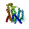
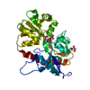
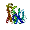
- PDB-1ypn: REDUCED FORM HYDROXYMETHYLBILANE SYNTHASE (K59Q MUTANT) CRYSTAL S... -
+ Open data

- Basic information
| Entry | Database: PDB / ID: 1ypn | ||||||
|---|---|---|---|---|---|---|---|
| Title | REDUCED FORM HYDROXYMETHYLBILANE SYNTHASE (K59Q MUTANT) CRYSTAL STRUCTURE AFTER 2 HOURS IN A FLOW CELL DETERMINED BY TIME-RESOLVED LAUE DIFFRACTION | ||||||
 Components Components | HYDROXYMETHYLBILANE SYNTHASE | ||||||
 Keywords Keywords | TRANSFERASE / BIOSYNTHESIS OF LINEAR TETRAPYRROLE / ALL ALPHA/BETA | ||||||
| Function / homology |  Function and homology information Function and homology informationtetrapyrrole biosynthetic process / hydroxymethylbilane synthase / hydroxymethylbilane synthase activity / : / heme biosynthetic process / cytoplasm / cytosol Similarity search - Function | ||||||
| Biological species |  | ||||||
| Method |  X-RAY DIFFRACTION / SYNCHROTRON / MOLECULAR REPLACEMENT / Resolution: 2.3 Å X-RAY DIFFRACTION / SYNCHROTRON / MOLECULAR REPLACEMENT / Resolution: 2.3 Å | ||||||
 Authors Authors | Helliwell, J.R. / Nieh, Y.P. / Raftery, J. / Cassetta, A. / Habash, J. / Carr, P.D. / Ursby, T. / Wulff, M. / Thompson, A.W. / Niemann, A.C. / Haedener, A. | ||||||
 Citation Citation | Journal: J.Chem.Soc.,Faraday Trans. / Year: 1998 Title: Time-Resolved Structures of Hydroxymethylbilane Synthase (Lys59Gln Mutant) as It Isloaded with Substrate in the Crystal Determined by Laue Diffraction Authors: Helliwell, J.R. / Nieh, Y.P. / Cassetta, A. / Habash, J. / Carr, P.D. / Ursby, T. / Wulff, M. / Thompson, A.W. / Niemann, A.C. / Haedener, A. #1: Journal: Time-Resolved Diffraction (in: Oxford Series on Synchrotron Radiation, V.2)Year: 1997 Title: Time-Resolved Protein Crystal Diffraction: Determination by the Laue Method of the Behaviour of the Enzyme Hydroxymethylbilane Synthase (Lys59Gln Mutant) as It is Loaded with Substrate in the Crystal Authors: Helliwell, J.R. / Nieh, Y.P. / Cassetta, A. / Raftery, J. / Haedener, A. / Niemann, A.C. / Battersby, A.R. / Carr, P.D. / Wulff, M. / Ursby, T. / Moy, J.P. / Thompson, A.W. | ||||||
| History |
|
- Structure visualization
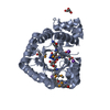
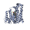
| Structure viewer | Molecule: MolmilJmol/JSmol |
|---|
- Downloads & links
-Download
| PDBx/mmCIF format | 1ypn.cif.gz | 68.9 KB | Display | PDBx/mmCIF format |
|---|---|---|---|---|
| PDB format | pdb1ypn.ent.gz | 53.5 KB | Display | PDB format |
| PDBx/mmJSON format | 1ypn.json.gz | Tree view | PDBx/mmJSON format | |
| Others |  Other downloads Other downloads |
-Validation report
| Arichive directory | https://data.pdbj.org/pub/pdb/validation_reports/yp/1ypnftp://data.pdbj.org/pub/pdb/validation_reports/yp/1ypn | HTTPS FTP |
|---|
-Related structure data
| Related structure data |  1ah5S S: Starting model for refinement |
|---|---|
| Similar structure data |


-Links
 PDBj
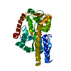
PDBj- Assembly
| Deposited unit | 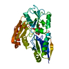
| ||||||||
|---|---|---|---|---|---|---|---|---|---|
| 1 |
| ||||||||
| Unit cell |
|
-Components
| #1: Protein | Mass: 33890.754 Da / Num. of mol.: 1 / Fragment: THREE DOMAINS / Mutation: K59Q Source method: isolated from a genetically manipulated source Details: CONTAINS A DIPYRROMETHANE COFACTOR LINKED TO CYSTEINE 242 Source: (gene. exp.) |
|---|---|
| #2: Chemical | ChemComp-DPM /   Mass: 420.413 Da / Num. of mol.: 1 / Source method: obtained synthetically / Formula: C20H24N2O8 Mass: 420.413 Da / Num. of mol.: 1 / Source method: obtained synthetically / Formula: C20H24N2O8 |
| #3: Water | ChemComp-HOH /  Mass: 18.015 Da / Num. of mol.: 103 / Source method: isolated from a natural source / Formula: H2O Mass: 18.015 Da / Num. of mol.: 103 / Source method: isolated from a natural source / Formula: H2O |
-Experimental details
-Experiment
| Experiment | Method: X-RAY DIFFRACTION / Number of used crystals: 1 |
|---|
- Sample preparation
| Crystal | Density Matthews: 2.52 Å3/Da / Density % sol: 50 % |
|---|---|
| Crystal grow | pH: 5.3 / Details: pH 5.3 |
| Crystal grow | *PLUS Method: unknown |
-Data collection
| Diffraction | Mean temperature: 293 K |
|---|---|
| Diffraction source | Source: SYNCHROTRON / Site: ESRF  / Beamline: ID09 / Wavelength: 0.4 / Beamline: ID09 / Wavelength: 0.4 |
| Detector | Detector: CCD / Date: Nov 1, 1995 |
| Radiation | Monochromatic (M) / Laue (L): L / Scattering type: x-ray |
| Radiation wavelength | Wavelength: 0.4 Å / Relative weight: 1 |
| Reflection | Resolution: 2.3→14.2 Å / Num. obs: 11383 / % possible obs: 72.9 % / Redundancy: 4.5 % / Rmerge(I) obs: 0.113 / Net I/σ(I): 4.9 |
| Reflection shell | Resolution: 2.3→2.36 Å / Redundancy: 2.8 % / Rmerge(I) obs: 0.262 / Mean I/σ(I) obs: 2.3 / % possible all: 69.5 |
| Reflection shell | *PLUS % possible obs: 69.5 % |
- Processing
| Software |
| ||||||||||||||||||||||||||||||||||||||||||||||||||||||||||||
|---|---|---|---|---|---|---|---|---|---|---|---|---|---|---|---|---|---|---|---|---|---|---|---|---|---|---|---|---|---|---|---|---|---|---|---|---|---|---|---|---|---|---|---|---|---|---|---|---|---|---|---|---|---|---|---|---|---|---|---|---|---|
| Refinement | Method to determine structure: MOLECULAR REPLACEMENT Starting model: PDB ENTRY 1AH5 Resolution: 2.3→8 Å / Cross valid method: THROUGHOUT / σ(F): 0 Details: THE FOLLOWING REPRESENTS TWO POSSIBLE INTERPRETATIONS FOR THE RESIDUAL DENSITY PEAKS NEAR THE ACTIVE SITE. THESE ATOMS ARE NOT INCLUDED IN THE REFINED ENTRY. ES C1 MPU 317 18.066 7.441 24. ...Details: THE FOLLOWING REPRESENTS TWO POSSIBLE INTERPRETATIONS FOR THE RESIDUAL DENSITY PEAKS NEAR THE ACTIVE SITE. THESE ATOMS ARE NOT INCLUDED IN THE REFINED ENTRY. ES C1 MPU 317 18.066 7.441 24.741 1.00 37.78 C2 MPU 317 18.656 7.598 25.957 1.00 34.85 C3 MPU 317 17.636 7.431 26.931 1.00 34.45 C4 MPU 317 16.462 7.194 26.266 1.00 34.64 C5 MPU 317 20.126 7.927 26.193 1.00 35.91 C6 MPU 317 20.580 9.306 25.663 1.00 37.14 C7 MPU 317 17.812 7.376 28.420 1.00 39.33 C8 MPU 317 17.441 8.637 29.164 1.00 41.42 C9 MPU 317 17.216 8.375 30.652 1.00 44.03 CH MPU 317 18.660 7.490 23.334 1.00 39.78 N1 MPU 317 16.714 7.196 24.934 1.00 35.83 O1 MPU 317 21.627 9.819 26.117 1.00 26.39 O2 MPU 317 19.882 9.874 24.789 1.00 39.95 O3 MPU 317 17.777 7.371 31.189 1.00 41.51 O4 MPU 317 16.457 9.168 31.272 1.00 44.53 N2 MPU 317 18.671 6.176 22.688 1.00 35.23 EP C1 MPU 317 16.744 7.168 25.091 1.00 53.28 C2 MPU 317 18.034 7.174 24.679 1.00 54.37 C3 MPU 317 18.838 7.176 25.840 1.00 52.21 C5 MPU 317 18.501 7.135 23.241 1.00 59.11 C6 MPU 317 18.419 8.470 22.530 1.00 64.68 C7 MPU 317 20.339 7.170 25.898 1.00 49.54 C8 MPU 317 20.913 8.218 26.812 1.00 42.08 C9 MPU 317 20.840 9.587 26.213 1.00 32.97 CH MPU 317 15.457 7.163 24.306 1.00 54.00 N1 MPU 317 16.724 7.163 26.458 1.00 55.05 O1 MPU 317 19.289 9.333 22.797 1.00 65.91 O2 MPU 317 17.479 8.660 21.719 1.00 63.89 O3 MPU 317 21.913 10.194 26.071 1.00 33.28 O4 MPU 317 19.729 10.043 25.874 1.00 30.65 C4 MPU 317 18.012 7.163 26.924 1.00 55.19 N2 MPU 317 14.560 8.204 24.757 1.00 50.26 C1 MPU 318 17.611 7.981 29.348 1.00 60.97 C2 MPU 318 17.362 9.319 29.304 1.00 63.79 C3 MPU 318 16.609 9.638 30.459 1.00 64.74 CH MPU 318 18.352 7.071 28.406 1.00 57.37 N1 MPU 318 17.042 7.467 30.482 1.00 61.70 C4 MPU 318 16.426 8.478 31.164 1.00 63.62 C1 MPU 319 15.396 9.357 33.385 1.00 60.58 C2 MPU 319 14.647 9.451 34.523 1.00 57.45 C3 MPU 319 14.739 10.792 34.976 1.00 58.19 CH MPU 319 15.679 8.177 32.460 1.00 61.22 N1 MPU 319 15.937 10.591 33.129 1.00 59.35 C4 MPU 319 15.537 11.460 34.095 1.00 57.90 C1 MPU 320 17.073 13.274 35.005 1.00 60.74 C2 MPU 320 17.015 13.824 36.250 1.00 61.26 C3 MPU 320 18.349 13.936 36.717 1.00 64.14 C4 MPU 320 19.163 13.453 35.734 1.00 63.20 CH MPU 320 15.988 12.900 34.027 1.00 58.80 N1 MPU 320 18.383 13.052 34.687 1.00 62.92 MPU 318,319,320 HAVE NO SIDE CHAINS MODELLED, DUE TO DISORDER/POOR ELECTRON DENSITY.
| ||||||||||||||||||||||||||||||||||||||||||||||||||||||||||||
| Displacement parameters | Biso mean: 22 Å2 | ||||||||||||||||||||||||||||||||||||||||||||||||||||||||||||
| Refinement step | Cycle: LAST / Resolution: 2.3→8 Å
| ||||||||||||||||||||||||||||||||||||||||||||||||||||||||||||
| Refine LS restraints |
| ||||||||||||||||||||||||||||||||||||||||||||||||||||||||||||
| LS refinement shell | Resolution: 2.3→2.4 Å / Total num. of bins used: 8
| ||||||||||||||||||||||||||||||||||||||||||||||||||||||||||||
| Xplor file |
| ||||||||||||||||||||||||||||||||||||||||||||||||||||||||||||
| Software | *PLUS Name: X-PLOR / Version: 3.1F / Classification: refinement | ||||||||||||||||||||||||||||||||||||||||||||||||||||||||||||
| Refine LS restraints | *PLUS
|
