 Movie
Movie Controller

[English] 日本語
 Yorodumi
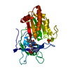
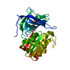
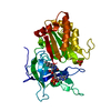
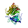
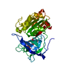
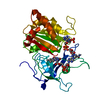
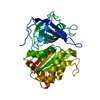
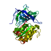
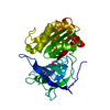
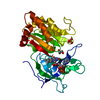
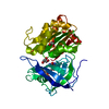
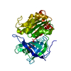
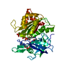
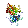
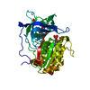
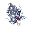
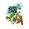
- PDB-1gr1: Structure of Ferredoxin-NADP+ Reductase with Glu 139 replaced by ... -
+ Open data

- Basic information
| Entry | Database: PDB / ID: 1gr1 | ||||||
|---|---|---|---|---|---|---|---|
| Title | Structure of Ferredoxin-NADP+ Reductase with Glu 139 replaced by Lys (E139K) | ||||||
 Components Components | FERREDOXIN--NADP+ REDUCTASE | ||||||
 Keywords Keywords | OXIDOREDUCTASE / FLAVOPROTEIN / FAD / FNR / NADP+ REDUCTASE | ||||||
| Function / homology |  Function and homology information Function and homology informationferredoxin-NADP+ reductase / ferredoxin-NADP+ reductase activity / phycobilisome / plasma membrane-derived thylakoid membrane / electron transport chain / NADP binding / flavin adenine dinucleotide binding Similarity search - Function | ||||||
| Biological species |  NOSTOC SP. PCC7119 (bacteria) NOSTOC SP. PCC7119 (bacteria) | ||||||
| Method |  X-RAY DIFFRACTION / MOLECULAR REPLACEMENT / Resolution: 2.5 Å X-RAY DIFFRACTION / MOLECULAR REPLACEMENT / Resolution: 2.5 Å | ||||||
 Authors Authors | Hermoso, J.A. / Mayoral, T. / Medina, M. / Sanz-Aparicio, J. / Gomez-Moreno, C. | ||||||
 Citation Citation | Journal: Eur.J.Biochem. / Year: 2002 Title: Probing the Role of Glutamic Acid 139 of Anabena Ferrodoxin-Nadp+ Reductase in the Interaction with Substrates Authors: Faro, M. / Frago, S. / Mayoral, T. / Hermoso, J.A. / Sanz-Aparico, J. / Gomez-Moreno, C. / Medina, M. #1: Journal: J.Mol.Biol. / Year: 1996Title: X-Ray Structure of the Ferredoxin:Nadp+ Reductase from the Cyanobacterium Anabanena Pcc 7119 at 1.8A Resolution, and Crystallographic Studies of Nadp Binding at 2.25A Resolution Authors: Serre, L. / Vellieux, F.M.D. / Medina, M. / Gomez-Moreno, C. / Fontecilla, J.C. / Frey, M. | ||||||
| History |
|
- Structure visualization
| Structure viewer | Molecule: MolmilJmol/JSmol |
|---|
- Downloads & links
-Download
| PDBx/mmCIF format | 1gr1.cif.gz | 79.9 KB | Display | PDBx/mmCIF format |
|---|---|---|---|---|
| PDB format | pdb1gr1.ent.gz | 57.8 KB | Display | PDB format |
| PDBx/mmJSON format | 1gr1.json.gz | Tree view | PDBx/mmJSON format | |
| Others |  Other downloads Other downloads |
-Validation report
| Arichive directory | https://data.pdbj.org/pub/pdb/validation_reports/gr/1gr1ftp://data.pdbj.org/pub/pdb/validation_reports/gr/1gr1 | HTTPS FTP |
|---|
-Related structure data
| Related structure data | 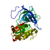 1queS S: Starting model for refinement |
|---|---|
| Similar structure data |


-Links
 PDBj
PDBj

- Assembly
| Deposited unit | 
| ||||||||
|---|---|---|---|---|---|---|---|---|---|
| 1 |
| ||||||||
| Unit cell |
|
-Components
| #1: Protein | Mass: 34041.742 Da / Num. of mol.: 1 / Mutation: YES Source method: isolated from a genetically manipulated source Source: (gene. exp.) NOSTOC SP. PCC7119 (bacteria) / Production host: |
|---|---|
| #2: Chemical | ChemComp-FAD /   Mass: 785.550 Da / Num. of mol.: 1 / Source method: obtained synthetically / Formula: C27H33N9O15P2 / Comment: FAD*YM Mass: 785.550 Da / Num. of mol.: 1 / Source method: obtained synthetically / Formula: C27H33N9O15P2 / Comment: FAD*YM |
| #3: Chemical | ChemComp-SO4 /   Mass: 96.063 Da / Num. of mol.: 1 / Source method: obtained synthetically / Formula: SO4 Mass: 96.063 Da / Num. of mol.: 1 / Source method: obtained synthetically / Formula: SO4 |
| #4: Water | ChemComp-HOH /  Mass: 18.015 Da / Num. of mol.: 203 / Source method: isolated from a natural source / Formula: H2O Mass: 18.015 Da / Num. of mol.: 203 / Source method: isolated from a natural source / Formula: H2O |
| Compound details | ENGINEERED |
-Experimental details
-Experiment
| Experiment | Method: X-RAY DIFFRACTION / Number of used crystals: 1 |
|---|
- Sample preparation
| Crystal | Density Matthews: 3.3 Å3/Da / Density % sol: 63 % | ||||||||||||||||||||||||||||||||||||||||||
|---|---|---|---|---|---|---|---|---|---|---|---|---|---|---|---|---|---|---|---|---|---|---|---|---|---|---|---|---|---|---|---|---|---|---|---|---|---|---|---|---|---|---|---|
| Crystal grow | pH: 5.5 / Details: pH 5.50 | ||||||||||||||||||||||||||||||||||||||||||
| Crystal grow | *PLUS Temperature: 20-23 ℃ / pH: 8 / Method: vapor diffusion, hanging drop | ||||||||||||||||||||||||||||||||||||||||||
| Components of the solutions | *PLUS
|
-Data collection
| Diffraction | Mean temperature: 100 K |
|---|---|
| Diffraction source | Source: ROTATING ANODE / Type: ENRAF-NONIUS FR571 / Wavelength: 1.5418 |
| Detector | Type: MARRESEARCH / Detector: IMAGE PLATE / Date: Jun 15, 2001 |
| Radiation | Monochromator: GRAPHITE / Protocol: SINGLE WAVELENGTH / Monochromatic (M) / Laue (L): M / Scattering type: x-ray |
| Radiation wavelength | Wavelength: 1.5418 Å / Relative weight: 1 |
| Reflection | Resolution: 2.5→27.3 Å / Num. obs: 13944 / % possible obs: 97.1 % / Observed criterion σ(I): 3 / Redundancy: 3.9 % / Rmerge(I) obs: 0.167 / Net I/σ(I): 3.2 |
| Reflection shell | Resolution: 2.5→2.64 Å / Redundancy: 4.1 % / Rmerge(I) obs: 0.264 / Mean I/σ(I) obs: 2.6 / % possible all: 99.9 |
| Reflection shell | *PLUS % possible obs: 99.9 % |
- Processing
| Software |
| ||||||||||||||||||||||||||||||||||||||||||||||||||||||||||||
|---|---|---|---|---|---|---|---|---|---|---|---|---|---|---|---|---|---|---|---|---|---|---|---|---|---|---|---|---|---|---|---|---|---|---|---|---|---|---|---|---|---|---|---|---|---|---|---|---|---|---|---|---|---|---|---|---|---|---|---|---|---|
| Refinement | Method to determine structure: MOLECULAR REPLACEMENT Starting model: PDB ENTRY 1QUE Resolution: 2.5→10 Å / Data cutoff high absF: 1000000 / Data cutoff low absF: 0.001 / Cross valid method: FREE R-VALUE / σ(F): 0 / Details: NO ELECTRON DENSITY PRESENT FOR RESIDUES 1-8
| ||||||||||||||||||||||||||||||||||||||||||||||||||||||||||||
| Displacement parameters | Biso mean: 19.15 Å2 | ||||||||||||||||||||||||||||||||||||||||||||||||||||||||||||
| Refinement step | Cycle: LAST / Resolution: 2.5→10 Å
| ||||||||||||||||||||||||||||||||||||||||||||||||||||||||||||
| Refine LS restraints |
| ||||||||||||||||||||||||||||||||||||||||||||||||||||||||||||
| LS refinement shell | Resolution: 2.5→2.61 Å / Total num. of bins used: 8
| ||||||||||||||||||||||||||||||||||||||||||||||||||||||||||||
| Refinement | *PLUS Lowest resolution: 10 Å | ||||||||||||||||||||||||||||||||||||||||||||||||||||||||||||
| Solvent computation | *PLUS | ||||||||||||||||||||||||||||||||||||||||||||||||||||||||||||
| Displacement parameters | *PLUS | ||||||||||||||||||||||||||||||||||||||||||||||||||||||||||||
| Refine LS restraints | *PLUS
|
