 Movie
Movie Controller

[English] 日本語
 Yorodumi
- PDB-1que: X-RAY STRUCTURE OF THE FERREDOXIN:NADP+ REDUCTASE FROM THE CYANOB... -
+ Open data

- Basic information
| Entry | Database: PDB / ID: 1que | ||||||
|---|---|---|---|---|---|---|---|
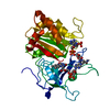
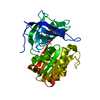
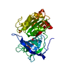
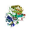
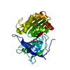
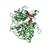
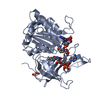
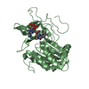
| Title | X-RAY STRUCTURE OF THE FERREDOXIN:NADP+ REDUCTASE FROM THE CYANOBACTERIUM ANABAENA PCC 7119 AT 1.8 ANGSTROMS | ||||||
 Components Components | FERREDOXIN--NADP+ REDUCTASE | ||||||
 Keywords Keywords | OXIDOREDUCTASE / FLAVOPROTEIN / NADP / FAD / THYLAKOID MEMBRANE / HYCOBILISOME / FNR / NADP+ REDUCTASE | ||||||
| Function / homology |  Function and homology information Function and homology informationferredoxin-NADP+ reductase / ferredoxin-NADP+ reductase activity / phycobilisome / plasma membrane-derived thylakoid membrane / electron transport chain / NADP binding / flavin adenine dinucleotide binding Similarity search - Function | ||||||
| Biological species |  Nostoc sp. (bacteria) Nostoc sp. (bacteria) | ||||||
| Method |  X-RAY DIFFRACTION / MIR / Resolution: 1.8 Å X-RAY DIFFRACTION / MIR / Resolution: 1.8 Å | ||||||
 Authors Authors | Serre, L. / Frey, M. / Vellieux, F.M.D. | ||||||
 Citation Citation | Journal: J.Mol.Biol. / Year: 1996 Title: X-ray structure of the ferredoxin:NADP+ reductase from the cyanobacterium Anabaena PCC 7119 at 1.8 A resolution, and crystallographic studies of NADP+ binding at 2.25 A resolution. Authors: Serre, L. / Vellieux, F.M. / Medina, M. / Gomez-Moreno, C. / Fontecilla-Camps, J.C. / Frey, M. #1: Journal: J.Mol.Biol. / Year: 1991Title: Crystals of Anabaena Pcc 7119 Ferredoxin-Nadp+ Reductase Authors: Serre, L. / Medina, M. / Gomez-Moreno, C. / Fontecilla-Camps, J.C. / Frey, M. #2: Journal: Nucleic Acids Res. / Year: 1990Title: Sequence of the Ferredoxin-Nadp(+)-Reductase Gene from Anabaena Pcc 7119 Authors: Fillat, M.F. / Bakker, H.A. / Weisbeek, P.J. | ||||||
| History |
|
- Structure visualization
| Structure viewer | Molecule: MolmilJmol/JSmol |
|---|
- Downloads & links
-Download
| PDBx/mmCIF format | 1que.cif.gz | 83 KB | Display | PDBx/mmCIF format |
|---|---|---|---|---|
| PDB format | pdb1que.ent.gz | 61.9 KB | Display | PDB format |
| PDBx/mmJSON format | 1que.json.gz | Tree view | PDBx/mmJSON format | |
| Others |  Other downloads Other downloads |
-Validation report
| Arichive directory | https://data.pdbj.org/pub/pdb/validation_reports/qu/1queftp://data.pdbj.org/pub/pdb/validation_reports/qu/1que | HTTPS FTP |
|---|
-Related structure data


-Links
 PDBj
PDBj

- Assembly
| Deposited unit | 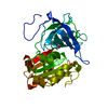
| ||||||||
|---|---|---|---|---|---|---|---|---|---|
| 1 |
| ||||||||
| Unit cell |
|
-Components
| #1: Protein | Mass: 34041.676 Da / Num. of mol.: 1 / Source method: isolated from a natural source / Source: (natural) Nostoc sp. (bacteria) / Strain: PCC 7119 / References: UniProt: P21890, ferredoxin-NADP+ reductase |
|---|---|
| #2: Chemical | ChemComp-SO4 /   Mass: 96.063 Da / Num. of mol.: 1 / Source method: obtained synthetically / Formula: SO4 Mass: 96.063 Da / Num. of mol.: 1 / Source method: obtained synthetically / Formula: SO4 |
| #3: Chemical | ChemComp-FAD /   Mass: 785.550 Da / Num. of mol.: 1 / Source method: obtained synthetically / Formula: C27H33N9O15P2 / Comment: FAD*YM Mass: 785.550 Da / Num. of mol.: 1 / Source method: obtained synthetically / Formula: C27H33N9O15P2 / Comment: FAD*YM |
| #4: Water | ChemComp-HOH /  Mass: 18.015 Da / Num. of mol.: 325 / Source method: isolated from a natural source / Formula: H2O Mass: 18.015 Da / Num. of mol.: 325 / Source method: isolated from a natural source / Formula: H2O |
-Experimental details
-Experiment
| Experiment | Method: X-RAY DIFFRACTION / Number of used crystals: 2 |
|---|
- Sample preparation
| Crystal | Density Matthews: 3 Å3/Da / Density % sol: 60 % | ||||||||||||||||||||||||||||||||||||||||||||||||||||||||||||
|---|---|---|---|---|---|---|---|---|---|---|---|---|---|---|---|---|---|---|---|---|---|---|---|---|---|---|---|---|---|---|---|---|---|---|---|---|---|---|---|---|---|---|---|---|---|---|---|---|---|---|---|---|---|---|---|---|---|---|---|---|---|
| Crystal grow | Method: vapor diffusion, hanging drop / pH: 7 Details: SEE REFERENCE 1., pH 7.0, vapor diffusion - hanging drop | ||||||||||||||||||||||||||||||||||||||||||||||||||||||||||||
| Crystal grow | *PLUS Temperature: 20 ℃ / Method: vapor diffusion, hanging drop | ||||||||||||||||||||||||||||||||||||||||||||||||||||||||||||
| Components of the solutions | *PLUS
|
-Data collection
| Diffraction | Mean temperature: 300 K |
|---|---|
| Diffraction source | Source: ROTATING ANODE / Type: RIGAKU RUH2R / Wavelength: 1.5418 |
| Detector | Type: SIEMENS / Detector: AREA DETECTOR / Date: Oct 1, 1992 |
| Radiation | Monochromator: SI(111) / Monochromatic (M) / Laue (L): M / Scattering type: x-ray |
| Radiation wavelength | Wavelength: 1.5418 Å / Relative weight: 1 |
| Reflection | Resolution: 2.1→20 Å / Num. obs: 20337 / % possible obs: 85 % / Observed criterion σ(I): 2 / Redundancy: 4 % / Rsym value: 0.07 |
| Reflection shell | Resolution: 2.1→2.2 Å / Redundancy: 3 % / % possible all: 34 |
| Reflection | *PLUS Lowest resolution: 20 Å / Num. measured all: 96286 / Rmerge(I) obs: 0.07 |
| Reflection shell | *PLUS % possible obs: 34 % |
- Processing
| Software |
| ||||||||||||||||||||||||||||||||||||||||||||||||||||||||||||||||||||||||||||||||
|---|---|---|---|---|---|---|---|---|---|---|---|---|---|---|---|---|---|---|---|---|---|---|---|---|---|---|---|---|---|---|---|---|---|---|---|---|---|---|---|---|---|---|---|---|---|---|---|---|---|---|---|---|---|---|---|---|---|---|---|---|---|---|---|---|---|---|---|---|---|---|---|---|---|---|---|---|---|---|---|---|---|
| Refinement | Method to determine structure: MIR / Resolution: 1.8→15 Å / Cross valid method: FREE-R / σ(F): 4 Details: THE INITIAL MODEL WAS BUILT IN A M.I.R SOLVENT FLATTENED ELECTRON DENSITY MAP AT 2.6 ANGSTROMS RESOLUTION WITH THE SPINACH FNR MODEL AS A GUIDE (KARPLUS, DANIELS, HERRIOTT, SCIENCE 1991, 251 ...Details: THE INITIAL MODEL WAS BUILT IN A M.I.R SOLVENT FLATTENED ELECTRON DENSITY MAP AT 2.6 ANGSTROMS RESOLUTION WITH THE SPINACH FNR MODEL AS A GUIDE (KARPLUS, DANIELS, HERRIOTT, SCIENCE 1991, 251 60-66). RESIDUES 1 - 8 AND 106 - 112 ARE POORLY DEFINED IN THE ELECTRON DENSITY MAP AND THEY HAVE BEEN TENTATIVELY MODELED. WATER MOLECULES HAVE BEEN NUMBERED ACCORDING THE INCREASING VALUES OF THEIR TEMPERATURE FACTORS STARTING WITH 401. WATER SITES CLOSER THAN 2.5 ANGSTROMS TO PROTEIN ATOMS OR OTHER WATER SITES REFLECTS, MOST PROBABLY, DISORDER.
| ||||||||||||||||||||||||||||||||||||||||||||||||||||||||||||||||||||||||||||||||
| Displacement parameters | Biso mean: 14.7 Å2 | ||||||||||||||||||||||||||||||||||||||||||||||||||||||||||||||||||||||||||||||||
| Refine analyze | Luzzati coordinate error obs: 0.2 Å / Luzzati d res low obs: 15 Å | ||||||||||||||||||||||||||||||||||||||||||||||||||||||||||||||||||||||||||||||||
| Refinement step | Cycle: LAST / Resolution: 1.8→15 Å
| ||||||||||||||||||||||||||||||||||||||||||||||||||||||||||||||||||||||||||||||||
| Refine LS restraints |
| ||||||||||||||||||||||||||||||||||||||||||||||||||||||||||||||||||||||||||||||||
| LS refinement shell | Resolution: 1.8→1.82 Å
| ||||||||||||||||||||||||||||||||||||||||||||||||||||||||||||||||||||||||||||||||
| Xplor file |
|
