[English] 日本語
Yorodumi
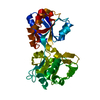
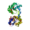
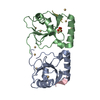
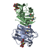
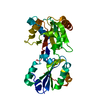
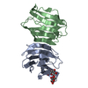
- PDB-1gpr: REFINED CRYSTAL STRUCTURE OF IIA DOMAIN OF THE GLUCOSE PERMEASE O... -

+
Open data


ID or keywords:

Loading...

-
Basic information

Entry
Database: PDB / ID: 1gpr
TitleREFINED CRYSTAL STRUCTURE OF IIA DOMAIN OF THE GLUCOSE PERMEASE OF BACILLUS SUBTILIS AT 1.9 ANGSTROMS RESOLUTION
ComponentsGLUCOSE PERMEASE
KeywordsPHOSPHOTRANSFERASE
Function / homology
Function and homology information


protein-Npi-phosphohistidine-D-glucose phosphotransferase / protein-phosphocysteine-glucose phosphotransferase system transporter activity / protein-Npi-phosphohistidine-sugar phosphotransferase / protein-phosphocysteine-sugar phosphotransferase activity / protein-N(pi)-phosphohistidine--N-acetyl-D-glucosamine phosphotransferase activity / protein-N(PI)-phosphohistidine-sugar phosphotransferase activity / glucose transmembrane transporter activity / phosphoenolpyruvate-dependent sugar phosphotransferase system / kinase activity / phosphorylation / plasma membrane
Similarity search - Function
Phosphotransferase system, maltose/glucose-specific subfamily IIC component / PTS system glucose-specific IIBC component / Phosphotransferase system, IIB component, type 1 / Phosphotransferase system, EIIC component, type 1 / Phosphotransferase system EIIB, cysteine phosphorylation site / Glucose permease domain IIB / phosphotransferase system, EIIB / PTS EIIB domains cysteine phosphorylation site signature. / PTS_EIIB type-1 domain profile. / PTS_EIIC type-1 domain profile. ...Phosphotransferase system, maltose/glucose-specific subfamily IIC component / PTS system glucose-specific IIBC component / Phosphotransferase system, IIB component, type 1 / Phosphotransferase system, EIIC component, type 1 / Phosphotransferase system EIIB, cysteine phosphorylation site / Glucose permease domain IIB / phosphotransferase system, EIIB / PTS EIIB domains cysteine phosphorylation site signature. / PTS_EIIB type-1 domain profile. / PTS_EIIC type-1 domain profile. / Phosphotransferase system, EIIC / Phosphotransferase system, EIIC / PTS EIIA domains phosphorylation site signature 1. / Phosphotransferase system, sugar-specific permease EIIA type 1 / phosphoenolpyruvate-dependent sugar phosphotransferase system, EIIA 1 / PTS_EIIA type-1 domain profile. / Glucose Permease (Domain IIA) / Glucose Permease (Domain IIA) / Duplicated hybrid motif / Distorted Sandwich / Mainly Beta
Similarity search - Domain/homology
PTS system glucose-specific EIICBA component / Protein-N(Pi)-phosphohistidine-sugar phosphotransferase
Similarity search - Component
Biological speciesBacillus subtilis (bacteria)
MethodX-RAY DIFFRACTION / Resolution: 1.9 Å
AuthorsLiao, D.-I. / Herzberg, O.
Citation
Journal: J.Biol.Chem. / Year: 1992
Title: An atomic model for protein-protein phosphoryl group transfer.
Authors: Herzberg, O.
#1: Journal: Biochemistry / Year: 1991
Title: Structure of the Iia Domain of the Glucose Permease of Bacillus Subtilis at 2.2 Angstroms Resolution
Authors: Liao, D.-I. / Kapadia, G. / Reddy, P. / Saier Junior, M.H. / Reizer, J. / Herzberg, O.
#2: Journal: J.Mol.Biol. / Year: 1991
Title: Crystallization of the Iia Domain of the Glucose Permease of Bacillus Subtilis
Authors: Kapadia, G. / Chen, C.C.H. / Reddy, P. / Saier Junior, M.H. / Reizer, J. / Herzberg, O.
#3: Journal: Proc.Natl.Acad.Sci.USA / Year: 1991
Title: Three-Dimensional Structure of the Escherichia Coli Phosphocarrier Protein IIIGlc
Authors: Worthylake, D. / Meadow, N.D. / Roseman, S. / Liao, D.-I. / Herzberg, O. / Remington, S.J.
History
DepositionSep 25, 1991Processing site: BNL
Revision 1.0Oct 31, 1993Provider: repository / Type: Initial release
Revision 1.1Mar 24, 2008Group: Version format compliance
Revision 1.2Jul 13, 2011Group: Version format compliance
Revision 1.3Feb 7, 2024Group: Data collection / Database references / Other
Category: chem_comp_atom / chem_comp_bond ...chem_comp_atom / chem_comp_bond / database_2 / pdbx_database_status
Item: _database_2.pdbx_DOI / _database_2.pdbx_database_accession / _pdbx_database_status.process_site

-
Structure visualization

Structure viewerMolecule:
MolmilJmol/JSmol

Downloads & links

-
Assembly

Deposited unit
A: GLUCOSE PERMEASE


Theoretical massNumber of molelcules
Total (without water)17,3971
Polymers17,3971
Non-polymers00
Water1,802100
1
A: GLUCOSE PERMEASE

A: GLUCOSE PERMEASE


Theoretical massNumber of molelcules
Total (without water)34,7942
Polymers34,7942
Non-polymers00
Water362
TypeNameSymmetry operationNumber
identity operation1_555x,y,z1
crystal symmetry operation4_556x,-y,-z+11
Unit cell
Length a, b, c (Å)74.220, 54.940, 66.960
Angle α, β, γ (deg.)90.00, 90.00, 90.00
Int Tables number20
Space group name H-MC2221
Components on special symmetry positions
IDModelComponents
11A-1008-

HOH

21A-1033-

HOH

-
Components

#1: Protein GLUCOSE PERMEASE


Mass: 17396.787 Da / Num. of mol.: 1
Source method: isolated from a genetically manipulated source
Source: (gene. exp.) Bacillus subtilis (bacteria)
References: UniProt: P20166, UniProt: Q59250*PLUS, protein-Npi-phosphohistidine-sugar phosphotransferase
#2: Water ChemComp-HOH / water


Mass: 18.015 Da / Num. of mol.: 100 / Source method: isolated from a natural source / Formula: H2O
Compound detailsTHE BACILLUS SUBTILIS GLUCOSE PERMEASE (ENZYME II) CONSISTS OF THREE DOMAINS IIA, IIB, AND IIC, ...THE BACILLUS SUBTILIS GLUCOSE PERMEASE (ENZYME II) CONSISTS OF THREE DOMAINS IIA, IIB, AND IIC, RESIDING ON A SINGLE POLYPEPTIDE CHAIN, WITH THE ORDER IICBA. THE RECOMBINANT IIA DOMAIN (162 AMINO ACID RESIDUES) IS A SOLUBLE PROTEIN. SEQUENTIAL NUMBERING OF THIS RECOMBINANT PROTEIN IS USED HERE. SECONDARY STRUCTURE ASSIGNMENT IS ACCORDING TO KABSCH AND SANDER (BIOPOLYMERS 22, 2577-2637, 1983) EXCEPT RESIDUES GLU 12 - SER 17 CLEARLY ADOPT A BETA STRAND CONFORMATION, BUT ARE NOT INVOLVED IN HYDROGEN BONDING WITH OTHER STRANDS, AND THUS ARE NOT IDENTIFIED BY THE DSSP PROGRAM. BRIDGES OF SINGLE RESIDUES ARE NOT INCLUDED IN THE ASSIGNMENT.

-
Experimental details

-
Experiment

ExperimentMethod: X-RAY DIFFRACTION

-
Sample preparation

CrystalDensity Matthews: 1.96 Å3/Da / Density % sol: 37.26 %
Crystal grow
*PLUS
Method: other / Details: Stock, J.B.,(1990) Nature, 344, 395.

-
Data collection

RadiationScattering type: x-ray
Radiation wavelengthRelative weight: 1

-
Processing

SoftwareName: TNT / Classification: refinement
RefinementRfactor obs: 0.156 / Highest resolution: 1.9 Å
Refinement stepCycle: LAST / Highest resolution: 1.9 Å
ProteinNucleic acidLigandSolventTotal
Num. atoms1191 0 0 100 1291
Refine LS restraints
Refine-IDTypeDev ideal
X-RAY DIFFRACTIONt_bond_d0.021
X-RAY DIFFRACTIONt_angle_deg3.4
X-RAY DIFFRACTIONt_dihedral_angle_d
X-RAY DIFFRACTIONt_incorr_chiral_ct
X-RAY DIFFRACTIONt_pseud_angle
X-RAY DIFFRACTIONt_trig_c_planes
X-RAY DIFFRACTIONt_gen_planes
X-RAY DIFFRACTIONt_it
X-RAY DIFFRACTIONt_nbd
Refinement
*PLUS
Rfactor obs: 0.156 / Rfactor Rwork: 0.155
Solvent computation
*PLUS
Displacement parameters
*PLUS

+
About Yorodumi

-
News

-
Feb 9, 2022. New format data for meta-information of EMDB entries

New format data for meta-information of EMDB entries

  • Version 3 of the EMDB header file is now the official format.
  • The previous official version 1.9 will be removed from the archive.

Related info.:EMDB header

External links:wwPDB to switch to version 3 of the EMDB data model

-
Aug 12, 2020. Covid-19 info

Covid-19 info

URL: https://pdbjlvh1.pdbj.org/emnavi/covid19.php

New page: Covid-19 featured information page in EM Navigator.

Related info.:Covid-19 info / Mar 5, 2020. Novel coronavirus structure data

+
Mar 5, 2020. Novel coronavirus structure data

Novel coronavirus structure data

Related info.:Yorodumi Speices / Aug 12, 2020. Covid-19 info

External links:COVID-19 featured content - PDBj / Molecule of the Month (242):Coronavirus Proteases

+
Jan 31, 2019. EMDB accession codes are about to change! (news from PDBe EMDB page)

EMDB accession codes are about to change! (news from PDBe EMDB page)

  • The allocation of 4 digits for EMDB accession codes will soon come to an end. Whilst these codes will remain in use, new EMDB accession codes will include an additional digit and will expand incrementally as the available range of codes is exhausted. The current 4-digit format prefixed with “EMD-” (i.e. EMD-XXXX) will advance to a 5-digit format (i.e. EMD-XXXXX), and so on. It is currently estimated that the 4-digit codes will be depleted around Spring 2019, at which point the 5-digit format will come into force.
  • The EM Navigator/Yorodumi systems omit the EMD- prefix.

Related info.:Q: What is EMD? / ID/Accession-code notation in Yorodumi/EM Navigator

External links:EMDB Accession Codes are Changing Soon! / Contact to PDBj

+
Jul 12, 2017. Major update of PDB

Major update of PDB

  • wwPDB released updated PDB data conforming to the new PDBx/mmCIF dictionary.
  • This is a major update changing the version number from 4 to 5, and with Remediation, in which all the entries are updated.
  • In this update, many items about electron microscopy experimental information are reorganized (e.g. em_software).
  • Now, EM Navigator and Yorodumi are based on the updated data.

External links:wwPDB Remediation / Enriched Model Files Conforming to OneDep Data Standards Now Available in the PDB FTP Archive

-
Yorodumi

Thousand views of thousand structures

  • Yorodumi is a browser for structure data from EMDB, PDB, SASBDB, etc.
  • This page is also the successor to EM Navigator detail page, and also detail information page/front-end page for Omokage search.
  • The word "yorodu" (or yorozu) is an old Japanese word meaning "ten thousand". "mi" (miru) is to see.

Related info.:EMDB / PDB / SASBDB / Comparison of 3 databanks / Yorodumi Search / Aug 31, 2016. New EM Navigator & Yorodumi / Yorodumi Papers / Jmol/JSmol / Function and homology information / Changes in new EM Navigator and Yorodumi

Read more