 Movie
Movie Controller

[English] 日本語
 Yorodumi
- PDB-5xxr: Crystal structure of selenomethionine labelled RIBT from Bacillus... -
+ Open data

- Basic information
| Entry | Database: PDB / ID: 5xxr | ||||||
|---|---|---|---|---|---|---|---|
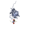
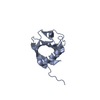
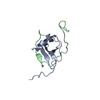
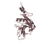
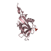
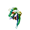
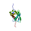
| Title | Crystal structure of selenomethionine labelled RIBT from Bacillus subtilis | ||||||
 Components Components | Protein RibT | ||||||
 Keywords Keywords | TRANSFERASE / Riboflavin / CoA / GNAT / acetylation | ||||||
| Function / homology |  Function and homology information Function and homology informationriboflavin biosynthetic process / acyltransferase activity, transferring groups other than amino-acyl groups / acyltransferase activity / Transferases; Acyltransferases; Transferring groups other than aminoacyl groups Similarity search - Function | ||||||
| Biological species |  | ||||||
| Method |  X-RAY DIFFRACTION / SYNCHROTRON / SAD / Resolution: 2.647 Å X-RAY DIFFRACTION / SYNCHROTRON / SAD / Resolution: 2.647 Å | ||||||
 Authors Authors | Srivastava, R. / Karthikeyan, S. | ||||||
| Funding support |  India, 1items India, 1items
| ||||||
 Citation Citation | Journal: J. Struct. Biol. / Year: 2018 Title: Structural characterization of ribT from Bacillus subtilis reveals it as a GCN5-related N-acetyltransferase. Authors: Srivastava, R. / Kaur, A. / Sharma, C. / Karthikeyan, S. | ||||||
| History |
|
- Structure visualization
| Structure viewer | Molecule: MolmilJmol/JSmol |
|---|
- Downloads & links
-Download
| PDBx/mmCIF format | 5xxr.cif.gz | 64.5 KB | Display | PDBx/mmCIF format |
|---|---|---|---|---|
| PDB format | pdb5xxr.ent.gz | 46.4 KB | Display | PDB format |
| PDBx/mmJSON format | 5xxr.json.gz | Tree view | PDBx/mmJSON format | |
| Others |  Other downloads Other downloads |
-Validation report
| Arichive directory | https://data.pdbj.org/pub/pdb/validation_reports/xx/5xxrftp://data.pdbj.org/pub/pdb/validation_reports/xx/5xxr | HTTPS FTP |
|---|
-Related structure data


-Links
 PDBj
PDBj


- Assembly
| Deposited unit | 
| ||||||||
|---|---|---|---|---|---|---|---|---|---|
| 1 | 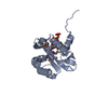
| ||||||||
| 2 | 
| ||||||||
| Unit cell |
|
-Components
| #1: Protein | Mass: 15900.222 Da / Num. of mol.: 2 Source method: isolated from a genetically manipulated source Source: (gene. exp.) Strain: 168 / Gene: ribT, BSU23240 / Plasmid: pET21b / Production host: References: UniProt: P17622, Transferases; Acyltransferases; Transferring groups other than aminoacyl groups #2: Chemical |   Mass: 767.534 Da / Num. of mol.: 2 / Source method: isolated from a natural source / Formula: C21H36N7O16P3S Mass: 767.534 Da / Num. of mol.: 2 / Source method: isolated from a natural source / Formula: C21H36N7O16P3S#3: Water | ChemComp-HOH / |  Mass: 18.015 Da / Num. of mol.: 23 / Source method: isolated from a natural source / Formula: H2O Mass: 18.015 Da / Num. of mol.: 23 / Source method: isolated from a natural source / Formula: H2OHas protein modification | Y | |
|---|
-Experimental details
-Experiment
| Experiment | Method: X-RAY DIFFRACTION / Number of used crystals: 1 |
|---|
- Sample preparation
| Crystal | Density Matthews: 2.17 Å3/Da / Density % sol: 43.46 % / Description: Two dimensional plate like |
|---|---|
| Crystal grow | Temperature: 293 K / Method: vapor diffusion, sitting drop / Details: 20% 2.7M sodium malonate, 80% 2.1M Malic acid / PH range: 7.0-7.5 |
-Data collection
| Diffraction | Mean temperature: 100 K |
|---|---|
| Diffraction source | Source: SYNCHROTRON / Site: ESRF  / Beamline: BM14 / Wavelength: 0.97883 Å / Beamline: BM14 / Wavelength: 0.97883 Å |
| Detector | Type: MARMOSAIC 225 mm CCD / Detector: CCD / Date: Nov 23, 2015 / Details: Mirror |
| Radiation | Monochromator: SI III / Protocol: SINGLE WAVELENGTH / Monochromatic (M) / Laue (L): M / Scattering type: x-ray |
| Radiation wavelength | Wavelength: 0.97883 Å / Relative weight: 1 |
| Reflection | Resolution: 2.63→50 Å / Num. obs: 6986 / % possible obs: 98.9 % / Observed criterion σ(I): -3 / Redundancy: 25.4 % / Biso Wilson estimate: 53 Å2 / CC1/2: 0.93 / Rmerge(I) obs: 0.191 / Net I/σ(I): 23.1 |
| Reflection shell | Resolution: 2.63→2.68 Å / Redundancy: 16.1 % / Rmerge(I) obs: 0.906 / Mean I/σ(I) obs: 1.6 / Num. unique obs: 315 / CC1/2: 0.68 / % possible all: 92.6 |
- Processing
| Software |
| ||||||||||||||||||||||||
|---|---|---|---|---|---|---|---|---|---|---|---|---|---|---|---|---|---|---|---|---|---|---|---|---|---|
| Refinement | Method to determine structure: SAD / Resolution: 2.647→36.544 Å / SU ML: 0.34 / Cross valid method: THROUGHOUT / σ(F): 1.34 / Phase error: 30.33 / Stereochemistry target values: ML
| ||||||||||||||||||||||||
| Solvent computation | Shrinkage radii: 0.9 Å / VDW probe radii: 1.11 Å / Solvent model: FLAT BULK SOLVENT MODEL | ||||||||||||||||||||||||
| Refinement step | Cycle: LAST / Resolution: 2.647→36.544 Å
| ||||||||||||||||||||||||
| Refine LS restraints |
| ||||||||||||||||||||||||
| LS refinement shell |
|
