 Movie
Movie Controller


+ Open data

- Basic information
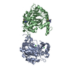
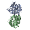
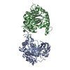
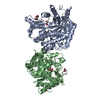
| Entry | Database: PDB / ID: 1gnx | |||||||||
|---|---|---|---|---|---|---|---|---|---|---|
| Title | b-glucosidase from Streptomyces sp | |||||||||
 Components Components | BETA-GLUCOSIDASE | |||||||||
 Keywords Keywords | HYDROLASE / GLYCOSYLTRANSFERASE / FAMILY 1 OF GLYCOSYL HYDROLASE | |||||||||
| Function / homology |  Function and homology information Function and homology informationbeta-glucosidase / beta-glucosidase activity / cellulose catabolic process / cytosol Similarity search - Function | |||||||||
| Biological species |  STREPTOMYCES SP. (bacteria) STREPTOMYCES SP. (bacteria) | |||||||||
| Method |  X-RAY DIFFRACTION / SYNCHROTRON / MOLECULAR REPLACEMENT / Resolution: 1.68 Å X-RAY DIFFRACTION / SYNCHROTRON / MOLECULAR REPLACEMENT / Resolution: 1.68 Å | |||||||||
 Authors Authors | Guasch, A. / Perez-Pons, J.A. / Vallmitjana, M. / Querol, E. / Coll, M. | |||||||||
 Citation Citation | Journal: To be Published Title: Beta-Glucosidase from Streptomyces Authors: Guasch, A. / Vallmitjana, M. / Perez, R. / Querol, E. / Perez-Pons, J.A. / Coll, M. #1: Journal: Acta Crystallogr.,Sect.D / Year: 2000 Title: Cloning, Overexpression, Crystallization and Preliminary X-Ray Analysis of a Family 1 Beta--Glucosidase from Streptomyces Authors: Guasch, A. / Vallmitjana, M. / Perez, R. / Querol, E. / Perez-Pons, J.A. / Coll, M. | |||||||||
| History |
| |||||||||
| Remark 700 | SHEET DETERMINATION METHOD: DSSP THE SHEETS PRESENTED AS "AB" IN EACH CHAIN ON SHEET RECORDS BELOW ... SHEET DETERMINATION METHOD: DSSP THE SHEETS PRESENTED AS "AB" IN EACH CHAIN ON SHEET RECORDS BELOW IS ACTUALLY AN 8-STRANDED BARREL THIS IS REPRESENTED BY A 9-STRANDED SHEET IN WHICH THE FIRST AND LAST STRANDS ARE IDENTICAL. SHEET DETERMINATION METHOD: DSSP THE SHEETS PRESENTED AS "BB" IN EACH CHAIN ON SHEET RECORDS BELOW IS ACTUALLY AN 8-STRANDED BARREL THIS IS REPRESENTED BY A 9-STRANDED SHEET IN WHICH THE FIRST AND LAST STRANDS ARE IDENTICAL. |
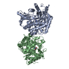
- Structure visualization
| Structure viewer | Molecule: MolmilJmol/JSmol |
|---|
- Downloads & links
-Download
| PDBx/mmCIF format | 1gnx.cif.gz | 196.3 KB | Display | PDBx/mmCIF format |
|---|---|---|---|---|
| PDB format | pdb1gnx.ent.gz | 154.8 KB | Display | PDB format |
| PDBx/mmJSON format | 1gnx.json.gz | Tree view | PDBx/mmJSON format | |
| Others |  Other downloads Other downloads |
-Validation report
| Arichive directory | https://data.pdbj.org/pub/pdb/validation_reports/gn/1gnxftp://data.pdbj.org/pub/pdb/validation_reports/gn/1gnx | HTTPS FTP |
|---|
-Related structure data
| Related structure data |  1cbgS S: Starting model for refinement |
|---|---|
| Similar structure data |


-Links
 PDBj
PDBj- Assembly
| Deposited unit | 
| ||||||||
|---|---|---|---|---|---|---|---|---|---|
| 1 |
| ||||||||
| Unit cell |
|
-Components
| #1: Protein | Mass: 52378.992 Da / Num. of mol.: 2 Source method: isolated from a genetically manipulated source Source: (gene. exp.) STREPTOMYCES SP. (bacteria) / Production host: #2: Polysaccharide | beta-D-fructofuranose-(2-1)-alpha-D-glucopyranose / sucrose |   Source method: isolated from a genetically manipulated source Details: oligosaccharide with reducing-end-to-reducing-end glycosidic bond References: sucrose #3: Chemical |   Mass: 96.063 Da / Num. of mol.: 3 / Source method: obtained synthetically / Formula: SO4 Mass: 96.063 Da / Num. of mol.: 3 / Source method: obtained synthetically / Formula: SO4#4: Water | ChemComp-HOH / |  Mass: 18.015 Da / Num. of mol.: 595 / Source method: isolated from a natural source / Formula: H2O Mass: 18.015 Da / Num. of mol.: 595 / Source method: isolated from a natural source / Formula: H2OCompound details | THE PROTEIN WAS SUBMITED TO LIMITED PROTEOLYSIS WITH TRYPSIN. THE MAJOR TRYPSIN-CLEAVAGE SITES ...THE PROTEIN WAS SUBMITED TO LIMITED PROTEOLYSI | |
|---|
-Experimental details
-Experiment
| Experiment | Method: X-RAY DIFFRACTION / Number of used crystals: 1 |
|---|
- Sample preparation
| Crystal | Density Matthews: 2.48 Å3/Da / Density % sol: 48.31 % |
|---|---|
| Crystal grow | pH: 7.5 / Details: 1.9 M AMMOMIUM SULFATE, 0.1 M HEPES PH 7.5 |
-Data collection
| Diffraction | Mean temperature: 103 K |
|---|---|
| Diffraction source | Source: SYNCHROTRON / Site: EMBL/DESY, HAMBURG  / Beamline: BW7A / Wavelength: 0.99 / Beamline: BW7A / Wavelength: 0.99 |
| Detector | Type: MARRESEARCH / Detector: IMAGE PLATE |
| Radiation | Protocol: SINGLE WAVELENGTH / Monochromatic (M) / Laue (L): M / Scattering type: x-ray |
| Radiation wavelength | Wavelength: 0.99 Å / Relative weight: 1 |
| Reflection | Resolution: 1.69→40 Å / Num. obs: 108818 / % possible obs: 97.7 % / Observed criterion σ(I): 0 / Redundancy: 85.6 % / Rmerge(I) obs: 0.038 / Net I/σ(I): 34.3 |
| Reflection shell | Resolution: 1.69→1.72 Å / Redundancy: 74.8 % / Rmerge(I) obs: 0.232 / Mean I/σ(I) obs: 5.86 / % possible all: 68.4 |
- Processing
| Software |
| ||||||||||||||||||||||||||||||||||||||||||||||||||||||||||||
|---|---|---|---|---|---|---|---|---|---|---|---|---|---|---|---|---|---|---|---|---|---|---|---|---|---|---|---|---|---|---|---|---|---|---|---|---|---|---|---|---|---|---|---|---|---|---|---|---|---|---|---|---|---|---|---|---|---|---|---|---|---|
| Refinement | Method to determine structure: MOLECULAR REPLACEMENT Starting model: PDB ENTRY 1CBG Resolution: 1.68→40 Å / Cross valid method: THROUGHOUT / σ(F): 0 / Stereochemistry target values: MLF
| ||||||||||||||||||||||||||||||||||||||||||||||||||||||||||||
| Displacement parameters | Biso mean: 81.4 Å2
| ||||||||||||||||||||||||||||||||||||||||||||||||||||||||||||
| Refinement step | Cycle: LAST / Resolution: 1.68→40 Å
| ||||||||||||||||||||||||||||||||||||||||||||||||||||||||||||
| Refine LS restraints |
|
