1-phosphatidylinositol binding / spectrin-associated cytoskeleton / regulation of intestinal absorption / actomyosin structure organization / cortical actin cytoskeleton organization / Neurexins and neuroligins / spectrin binding / cortical cytoskeleton / regulation of calcium ion transport / intercellular bridge ...1-phosphatidylinositol binding / spectrin-associated cytoskeleton / regulation of intestinal absorption / actomyosin structure organization / cortical actin cytoskeleton organization / Neurexins and neuroligins / spectrin binding / cortical cytoskeleton / regulation of calcium ion transport / intercellular bridge / positive regulation of protein localization to cell cortex / phosphoprotein binding / structural constituent of cytoskeleton / cell junction / mitotic spindle / actin cytoskeleton organization / actin binding / cell cortex / basolateral plasma membrane / cytoskeleton / calmodulin binding / nuclear body / cell division / protein-containing complex / plasma membrane / cytosol Similarity search - Function
Band 4.1 protein, chordates / SAB domain / Band 4.1, C-terminal / SAB domain / 4.1 protein C-terminal domain (CTD) / FERM adjacent (FA) / FERM adjacent (FA) / FERM adjacent (FA) / Acyl-CoA Binding Protein - #10 / Acyl-CoA Binding Protein ...Band 4.1 protein, chordates / SAB domain / Band 4.1, C-terminal / SAB domain / 4.1 protein C-terminal domain (CTD) / FERM adjacent (FA) / FERM adjacent (FA) / FERM adjacent (FA) / Acyl-CoA Binding Protein - #10 / Acyl-CoA Binding Protein / Ezrin/radixin/moesin-like / FERM, C-terminal PH-like domain / FERM C-terminal PH-like domain / FERM C-terminal PH-like domain / FERM, N-terminal / FERM N-terminal domain / FERM domain signature 1. / FERM conserved site / FERM domain signature 2. / FERM central domain / FERM/acyl-CoA-binding protein superfamily / Pleckstrin-homology domain (PH domain)/Phosphotyrosine-binding domain (PTB) / PH-domain like / FERM central domain / FERM superfamily, second domain / FERM domain / FERM domain profile. / Band 4.1 domain / Band 4.1 homologues / Phosphatidylinositol 3-kinase Catalytic Subunit; Chain A, domain 1 / Ubiquitin-like (UB roll) / PH-like domain superfamily / Roll / Ubiquitin-like domain superfamily / Roll / Up-down Bundle / Mainly Beta / Mainly Alpha / Alpha Beta Similarity search - Domain/homology
X-RAY DIFFRACTION / SYNCHROTRON / Resolution: 2.8 Å
Model details
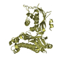
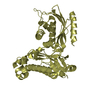
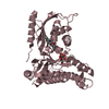
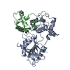
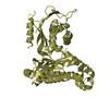
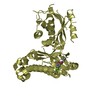
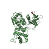
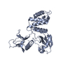
PROTEIN 4.1R 30KD N-TERMINAL DOMAIN PROVIDES MULTIPLE BINDING SITES FOR TRANSMEMBRANE PROTEINS AND ...PROTEIN 4.1R 30KD N-TERMINAL DOMAIN PROVIDES MULTIPLE BINDING SITES FOR TRANSMEMBRANE PROTEINS AND IS REGULATED BY CALMODULIN
In the structure databanks used in Yorodumi, some data are registered as the other names, "COVID-19 virus" and "2019-nCoV". Here are the details of the virus and the list of structure data.
Jan 31, 2019. EMDB accession codes are about to change! (news from PDBe EMDB page)
EMDB accession codes are about to change! (news from PDBe EMDB page)
The allocation of 4 digits for EMDB accession codes will soon come to an end. Whilst these codes will remain in use, new EMDB accession codes will include an additional digit and will expand incrementally as the available range of codes is exhausted. The current 4-digit format prefixed with “EMD-” (i.e. EMD-XXXX) will advance to a 5-digit format (i.e. EMD-XXXXX), and so on. It is currently estimated that the 4-digit codes will be depleted around Spring 2019, at which point the 5-digit format will come into force.
The EM Navigator/Yorodumi systems omit the EMD- prefix.
Related info.:Q: What is EMD? / ID/Accession-code notation in Yorodumi/EM Navigator
Yorodumi is a browser for structure data from EMDB, PDB, SASBDB, etc.
This page is also the successor to EM Navigator detail page, and also detail information page/front-end page for Omokage search.
The word "yorodu" (or yorozu) is an old Japanese word meaning "ten thousand". "mi" (miru) is to see.
Related info.:EMDB / PDB / SASBDB / Comparison of 3 databanks / Yorodumi Search / Aug 31, 2016. New EM Navigator & Yorodumi / Yorodumi Papers / Jmol/JSmol / Function and homology information / Changes in new EM Navigator and Yorodumi
 Movie
Movie Controller


 Open data
 Basic information
 Components
 Keywords
 Function and homology information
 Homo sapiens (human)
 X-RAY DIFFRACTION /
  Authors
 Citation
 Structure visualization
 Downloads & links
 Other downloads


 PDBj
PDBj


 Assembly
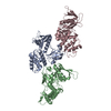


 Sample preparation
 / Beamline: 5.0.2 / Wavelength: 1
  Processing