 Movie
Movie Controller

[English] 日本語
 Yorodumi
- PDB-1ged: A positive charge route for the access of nadh to heme formed in ... -
+ Open data

- Basic information
| Entry | Database: PDB / ID: 1ged | ||||||
|---|---|---|---|---|---|---|---|
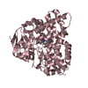
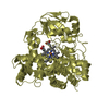
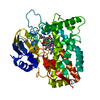
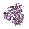
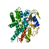
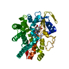
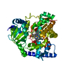
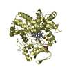
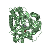
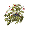
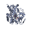
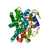
| Title | A positive charge route for the access of nadh to heme formed in the distal heme pocket of cytochrome p450nor | ||||||
 Components Components | CYTOCHROME P450 55A1 | ||||||
 Keywords Keywords | OXIDOREDUCTASE / Cytochrome P450nor / Nitric oxide reductase | ||||||
| Function / homology |  Function and homology information Function and homology informationnitric oxide reductase [NAD(P)+, nitrous oxide-forming] / nitric oxide reductase [NAD(P)H] activity / oxidoreductase activity, acting on paired donors, with incorporation or reduction of molecular oxygen / monooxygenase activity / iron ion binding / heme binding Similarity search - Function | ||||||
| Biological species |   Fusarium oxysporum (fungus) Fusarium oxysporum (fungus) | ||||||
| Method |  X-RAY DIFFRACTION / SYNCHROTRON / MOLECULAR REPLACEMENT / Resolution: 2 Å X-RAY DIFFRACTION / SYNCHROTRON / MOLECULAR REPLACEMENT / Resolution: 2 Å | ||||||
 Authors Authors | Kudo, T. / Takaya, N. / Park, S.-Y. / Shiro, Y. / Shoun, H. | ||||||
 Citation Citation | Journal: J.Biol.Chem. / Year: 2001 Title: A positively charged cluster formed in the heme-distal pocket of cytochrome P450nor is essential for interaction with NADH Authors: Kudo, T. / Takaya, N. / Park, S.-Y. / Shiro, Y. / Shoun, H. | ||||||
| History |
|
- Structure visualization
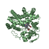
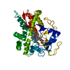
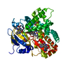
| Structure viewer | Molecule: MolmilJmol/JSmol |
|---|
- Downloads & links
-Download
| PDBx/mmCIF format | 1ged.cif.gz | 93.7 KB | Display | PDBx/mmCIF format |
|---|---|---|---|---|
| PDB format | pdb1ged.ent.gz | 69.7 KB | Display | PDB format |
| PDBx/mmJSON format | 1ged.json.gz | Tree view | PDBx/mmJSON format | |
| Others |  Other downloads Other downloads |
-Validation report
| Summary document | 1ged_validation.pdf.gz | 469.1 KB | Display | wwPDB validaton report |
|---|---|---|---|---|
| Full document | 1ged_full_validation.pdf.gz | 477.2 KB | Display | |
| Data in XML | 1ged_validation.xml.gz | 10.5 KB | Display | |
| Data in CIF | 1ged_validation.cif.gz | 15.8 KB | Display | |
| Arichive directory | https://data.pdbj.org/pub/pdb/validation_reports/ge/1gedftp://data.pdbj.org/pub/pdb/validation_reports/ge/1ged | HTTPS FTP |
-Related structure data
| Related structure data |  1romS S: Starting model for refinement |
|---|---|
| Similar structure data |


-Links
 PDBj
PDBj


- Assembly
| Deposited unit | 
| ||||||||
|---|---|---|---|---|---|---|---|---|---|
| 1 |
| ||||||||
| Unit cell |
|
-Components
| #1: Protein | Mass: 44420.691 Da / Num. of mol.: 1 Source method: isolated from a genetically manipulated source Source: (gene. exp.) Fusarium oxysporum (fungus) / Plasmid: PRSET-C / Production host:  References: UniProt: P23295, Oxidoreductases; Acting on paired donors, with incorporation or reduction of molecular oxygen | ||||
|---|---|---|---|---|---|
| #2: Chemical |   Mass: 79.904 Da / Num. of mol.: 2 / Source method: obtained synthetically / Formula: Br Mass: 79.904 Da / Num. of mol.: 2 / Source method: obtained synthetically / Formula: Br#3: Chemical | ChemComp-HEM / |   Mass: 616.487 Da / Num. of mol.: 1 / Source method: obtained synthetically / Formula: C34H32FeN4O4 Mass: 616.487 Da / Num. of mol.: 1 / Source method: obtained synthetically / Formula: C34H32FeN4O4#4: Water | ChemComp-HOH / |  Mass: 18.015 Da / Num. of mol.: 106 / Source method: isolated from a natural source / Formula: H2O Mass: 18.015 Da / Num. of mol.: 106 / Source method: isolated from a natural source / Formula: H2O |
-Experimental details
-Experiment
| Experiment | Method: X-RAY DIFFRACTION / Number of used crystals: 1 |
|---|
- Sample preparation
| Crystal | Density Matthews: 2.06 Å3/Da / Density % sol: 40.43 % | ||||||||||||||||||||||||||||||
|---|---|---|---|---|---|---|---|---|---|---|---|---|---|---|---|---|---|---|---|---|---|---|---|---|---|---|---|---|---|---|---|
| Crystal grow | Temperature: 298 K / Method: vapor diffusion, sitting drop / pH: 6.5 Details: PEG4000, pH 6.5, VAPOR DIFFUSION, SITTING DROP, temperature 298K | ||||||||||||||||||||||||||||||
| Crystal grow | *PLUS Temperature: 293 K / pH: 5.6 / Method: vapor diffusion | ||||||||||||||||||||||||||||||
| Components of the solutions | *PLUS
|
-Data collection
| Diffraction | Mean temperature: 100 K |
|---|---|
| Diffraction source | Source: SYNCHROTRON / Site: SPring-8  / Beamline: BL44B2 / Wavelength: 1 Å / Beamline: BL44B2 / Wavelength: 1 Å |
| Detector | Type: MAR CCD 165 mm / Detector: CCD / Date: Feb 20, 2000 / Details: Monochro. Si-111 |
| Radiation | Monochromator: Si-111 / Protocol: SINGLE WAVELENGTH / Monochromatic (M) / Laue (L): M / Scattering type: x-ray |
| Radiation wavelength | Wavelength: 1 Å / Relative weight: 1 |
| Reflection | Resolution: 2→30 Å / Num. all: 79264 / Num. obs: 23487 / % possible obs: 91.9 % / Observed criterion σ(F): 0 / Observed criterion σ(I): 0 / Redundancy: 3.4 % / Biso Wilson estimate: 19.9 Å2 / Rmerge(I) obs: 0.065 / Net I/σ(I): 10.5 |
| Reflection shell | Resolution: 2→2.09 Å / Redundancy: 3 % / Rmerge(I) obs: 0.305 / % possible all: 80.1 |
| Reflection | *PLUS Num. measured all: 79264 |
| Reflection shell | *PLUS % possible obs: 80.1 % |
- Processing
| Software |
| |||||||||||||||||||||||||
|---|---|---|---|---|---|---|---|---|---|---|---|---|---|---|---|---|---|---|---|---|---|---|---|---|---|---|
| Refinement | Method to determine structure: MOLECULAR REPLACEMENT Starting model: PDB ENTRY 1ROM Resolution: 2→30 Å / Isotropic thermal model: Overall / Cross valid method: THROUGHOUT / σ(F): 0 / σ(I): 0 / Stereochemistry target values: Engh & Huber
| |||||||||||||||||||||||||
| Displacement parameters | Biso mean: 35.8 Å2 | |||||||||||||||||||||||||
| Refine analyze | Luzzati coordinate error obs: 0.27 Å / Luzzati d res low obs: 5 Å / Luzzati sigma a obs: 0.35 Å | |||||||||||||||||||||||||
| Refinement step | Cycle: LAST / Resolution: 2→30 Å
| |||||||||||||||||||||||||
| Refine LS restraints |
| |||||||||||||||||||||||||
| LS refinement shell | Resolution: 2→2.13 Å / Rfactor Rfree error: 0.028
| |||||||||||||||||||||||||
| Software | *PLUS Name: X-PLOR / Version: 3.851 / Classification: refinement | |||||||||||||||||||||||||
| Refinement | *PLUS Highest resolution: 2 Å / Lowest resolution: 30 Å / σ(F): 0 / % reflection Rfree: 5 % / Rfactor obs: 0.21 | |||||||||||||||||||||||||
| Solvent computation | *PLUS | |||||||||||||||||||||||||
| Displacement parameters | *PLUS Biso mean: 35.8 Å2 | |||||||||||||||||||||||||
| Refine LS restraints | *PLUS
| |||||||||||||||||||||||||
| LS refinement shell | *PLUS Highest resolution: 2 Å / Rfactor Rfree: 0.346 / Rfactor Rwork: 0.315 |
