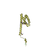
 Movie
Movie Controller


+ Open data

- Basic information
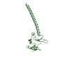
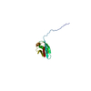
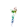
| Entry | Database: PDB / ID: 1gd2 | ||||||
|---|---|---|---|---|---|---|---|
| Title | CRYSTAL STRUCTURE OF BZIP TRANSCRIPTION FACTOR PAP1 BOUND TO DNA | ||||||
 Components Components |
| ||||||
 Keywords Keywords | transcription/DNA / basic leucine zipper / protein-DNA complex / transcription-DNA COMPLEX | ||||||
| Function / homology |  Function and homology information Function and homology informationDNA binding, bending / nucleosome binding / RNA polymerase II transcription regulator complex / cellular response to oxidative stress / DNA-binding transcription activator activity, RNA polymerase II-specific / DNA-binding transcription factor activity, RNA polymerase II-specific / transcription cis-regulatory region binding / RNA polymerase II cis-regulatory region sequence-specific DNA binding / regulation of transcription by RNA polymerase II / chromatin ...DNA binding, bending / nucleosome binding / RNA polymerase II transcription regulator complex / cellular response to oxidative stress / DNA-binding transcription activator activity, RNA polymerase II-specific / DNA-binding transcription factor activity, RNA polymerase II-specific / transcription cis-regulatory region binding / RNA polymerase II cis-regulatory region sequence-specific DNA binding / regulation of transcription by RNA polymerase II / chromatin / positive regulation of transcription by RNA polymerase II / nucleus / cytoplasm / cytosol Similarity search - Function | ||||||
| Biological species |  | ||||||
| Method |  X-RAY DIFFRACTION / Resolution: 2 Å X-RAY DIFFRACTION / Resolution: 2 Å | ||||||
 Authors Authors | Fujii, Y. / Shimizu, T. / Toda, T. / Yanagida, M. / Hakoshima, T. | ||||||
 Citation Citation | Journal: Nat.Struct.Biol. / Year: 2000 Title: Structural basis for the diversity of DNA recognition by bZIP transcription factors. Authors: Fujii, Y. / Shimizu, T. / Toda, T. / Yanagida, M. / Hakoshima, T. #1: Journal: Acta Crystallogr.,Sect.D / Year: 1998Title: Crystallographic characterization of Pap1-DNA complex Authors: Fujii, Y. / Ohira, T. / Kyogoku, Y. / Toda, T. / Yanagida, M. / Hakoshima, T. | ||||||
| History |
|
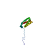
- Structure visualization
| Structure viewer | Molecule: MolmilJmol/JSmol |
|---|
- Downloads & links
-Download
| PDBx/mmCIF format | 1gd2.cif.gz | 119.5 KB | Display | PDBx/mmCIF format |
|---|---|---|---|---|
| PDB format | pdb1gd2.ent.gz | 90.5 KB | Display | PDB format |
| PDBx/mmJSON format | 1gd2.json.gz | Tree view | PDBx/mmJSON format | |
| Others |  Other downloads Other downloads |
-Validation report
| Arichive directory | https://data.pdbj.org/pub/pdb/validation_reports/gd/1gd2ftp://data.pdbj.org/pub/pdb/validation_reports/gd/1gd2 | HTTPS FTP |
|---|
-Related structure data
| Similar structure data |
|---|


-Links
 PDBj
PDBj


- Assembly
| Deposited unit | 
| ||||||||||
|---|---|---|---|---|---|---|---|---|---|---|---|
| 1 | 
| ||||||||||
| 2 | 
| ||||||||||
| 3 | 
| ||||||||||
| Unit cell |
| ||||||||||
| Details | The biological assmbly is a dimer constructed from chain E and F, chain G and H, and chain I and J. |
-Components
| #1: DNA chain | Mass: 3975.611 Da / Num. of mol.: 4 / Source method: obtained synthetically #2: Protein | Mass: 8357.505 Da / Num. of mol.: 6 / Fragment: LEUCINE ZIPPER DOMAIN Source method: isolated from a genetically manipulated source Source: (gene. exp.) Plasmid: PET3A / Production host:  #3: Water | ChemComp-HOH / |  Mass: 18.015 Da / Num. of mol.: 831 / Source method: isolated from a natural source / Formula: H2O Mass: 18.015 Da / Num. of mol.: 831 / Source method: isolated from a natural source / Formula: H2O |
|---|
-Experimental details
-Experiment
| Experiment | Method: X-RAY DIFFRACTION / Number of used crystals: 1 |
|---|
- Sample preparation
| Crystal | Density Matthews: 3.67 Å3/Da / Density % sol: 66.52 % | ||||||||||||||||||||
|---|---|---|---|---|---|---|---|---|---|---|---|---|---|---|---|---|---|---|---|---|---|
| Crystal grow | Temperature: 277 K / Method: vapor diffusion, hanging drop / pH: 5.8 Details: PEG 6000, KCl, MES, pH 5.8, VAPOR DIFFUSION, HANGING DROP, temperature 277K | ||||||||||||||||||||
| Components of the solutions |
| ||||||||||||||||||||
| Crystal grow | *PLUS Method: vapor diffusion | ||||||||||||||||||||
| Components of the solutions | *PLUS
|
-Data collection
| Diffraction | Mean temperature: 100 K |
|---|---|
| Diffraction source | Source: ROTATING ANODE / Type: RIGAKU FR-C / Wavelength: 1.5418 |
| Detector | Type: RIGAKU RAXIS IV++ / Detector: IMAGE PLATE / Date: May 12, 1997 |
| Radiation | Protocol: SINGLE WAVELENGTH / Monochromatic (M) / Laue (L): M / Scattering type: x-ray |
| Radiation wavelength | Wavelength: 1.5418 Å / Relative weight: 1 |
| Reflection | Resolution: 2→45.6 Å / Num. all: 127588 / Num. obs: 53501 / % possible obs: 83.4 % / Observed criterion σ(F): 2 / Observed criterion σ(I): 1 / Redundancy: 2.4 % / Biso Wilson estimate: 27 Å2 / Rmerge(I) obs: 0.052 / Net I/σ(I): 10.3 |
| Reflection shell | Resolution: 2→2.07 Å / Redundancy: 1.7 % / Rmerge(I) obs: 0.244 / Num. unique all: 3856 / % possible all: 60.2 |
| Reflection shell | *PLUS % possible obs: 60.2 % |
- Processing
| Software |
| |||||||||||||||||||||
|---|---|---|---|---|---|---|---|---|---|---|---|---|---|---|---|---|---|---|---|---|---|---|
| Refinement | Resolution: 2→20 Å / Cross valid method: THROUGHOUT / σ(F): 2 / σ(I): 1 / Stereochemistry target values: Engh & Huber
| |||||||||||||||||||||
| Refinement step | Cycle: LAST / Resolution: 2→20 Å
| |||||||||||||||||||||
| Refine LS restraints |
| |||||||||||||||||||||
| Software | *PLUS Name: CNS / Version: 0.9 / Classification: refinement | |||||||||||||||||||||
| Refinement | *PLUS | |||||||||||||||||||||
| Solvent computation | *PLUS | |||||||||||||||||||||
| Displacement parameters | *PLUS Biso mean: 42.8 Å2 | |||||||||||||||||||||
| Refine LS restraints | *PLUS
|
