 Movie
Movie Controller


+ Open data

- Basic information
| Entry | Database: PDB / ID: 1gcq | ||||||
|---|---|---|---|---|---|---|---|
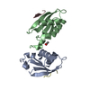
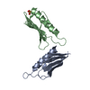
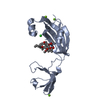
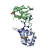
| Title | CRYSTAL STRUCTURE OF VAV AND GRB2 SH3 DOMAINS | ||||||
 Components Components |
| ||||||
 Keywords Keywords | SIGNALING PROTEIN/SIGNALING PROTEIN / SH3 domain / PROTEIN-PROTEIN COMPLEX / Grb2 / Vav / SIGNALING PROTEIN-SIGNALING PROTEIN COMPLEX | ||||||
| Function / homology |  Function and homology information Function and homology informationAzathioprine ADME / CD28 dependent Vav1 pathway / Erythropoietin activates RAS / RAC2 GTPase cycle / GPVI-mediated activation cascade / FCERI mediated MAPK activation / NRAGE signals death through JNK / Signaling by SCF-KIT / guanyl-nucleotide exchange factor adaptor activity / VEGFR2 mediated vascular permeability ...Azathioprine ADME / CD28 dependent Vav1 pathway / Erythropoietin activates RAS / RAC2 GTPase cycle / GPVI-mediated activation cascade / FCERI mediated MAPK activation / NRAGE signals death through JNK / Signaling by SCF-KIT / guanyl-nucleotide exchange factor adaptor activity / VEGFR2 mediated vascular permeability / RAC1 GTPase cycle / Grb2-EGFR complex / immune response-regulating cell surface receptor signaling pathway / FCERI mediated Ca+2 mobilization / G alpha (12/13) signalling events / PIP3 activates AKT signaling / RHOA GTPase cycle / Antigen activates B Cell Receptor (BCR) leading to generation of second messengers / RHOG GTPase cycle / Regulation of signaling by CBL / phosphorylation-dependent protein binding / Regulation of actin dynamics for phagocytic cup formation / PI5P, PP2A and IER3 Regulate PI3K/AKT Signaling / Interleukin-3, Interleukin-5 and GM-CSF signaling / branching involved in labyrinthine layer morphogenesis / STAT5 Activation / Co-inhibition by BTLA / neurotrophin TRKA receptor binding / COP9 signalosome / VEGFA-VEGFR2 Pathway / Activated NTRK2 signals through PI3K / MET receptor recycling / transmembrane receptor protein tyrosine kinase adaptor activity / Interleukin-15 signaling / positive regulation of natural killer cell mediated cytotoxicity / negative regulation of natural killer cell mediated cytotoxicity / Signaling by cytosolic FGFR1 fusion mutants / MET activates PTPN11 / natural killer cell activation / MET activates RAP1 and RAC1 / vesicle membrane / CD28 dependent Vav1 pathway / Signaling by LTK / Signal regulatory protein family interactions / MET activates PI3K/AKT signaling / Regulation of KIT signaling / epidermal growth factor receptor binding / natural killer cell mediated cytotoxicity / STAT5 activation downstream of FLT3 ITD mutants / PI-3K cascade:FGFR3 / small GTPase-mediated signal transduction / PI-3K cascade:FGFR2 / PI-3K cascade:FGFR4 / PI-3K cascade:FGFR1 / GRB2:SOS provides linkage to MAPK signaling for Integrins / endodermal cell differentiation / positive regulation of actin filament polymerization / RHOU GTPase cycle / B cell proliferation / regulation of MAPK cascade / RET signaling / T cell differentiation / Interleukin-3, Interleukin-5 and GM-CSF signaling / insulin receptor substrate binding / negative regulation of epidermal growth factor receptor signaling pathway / PI3K events in ERBB2 signaling / fibroblast growth factor receptor signaling pathway / PI3K Cascade / Role of LAT2/NTAL/LAB on calcium mobilization / Interleukin receptor SHC signaling / SOS-mediated signalling / Signal attenuation / Activated NTRK3 signals through RAS / Activated NTRK2 signals through RAS / GAB1 signalosome / SHC1 events in ERBB4 signaling / RHO GTPases Activate WASPs and WAVEs / Signalling to RAS / Schwann cell development / SHC-related events triggered by IGF1R / Activated NTRK2 signals through FRS2 and FRS3 / positive regulation of Rac protein signal transduction / SHC-mediated cascade:FGFR3 / phagocytosis / MET activates RAS signaling / ephrin receptor binding / SHC-mediated cascade:FGFR2 / SHC-mediated cascade:FGFR4 / Signaling by PDGFRA transmembrane, juxtamembrane and kinase domain mutants / Signaling by PDGFRA extracellular domain mutants / Erythropoietin activates RAS / SHC-mediated cascade:FGFR1 / Signaling by FGFR4 in disease / Signaling by CSF3 (G-CSF) / T cell costimulation / FRS-mediated FGFR3 signaling / Signaling by FLT3 ITD and TKD mutants / FRS-mediated FGFR2 signaling / FRS-mediated FGFR4 signaling / signal transduction in response to DNA damage Similarity search - Function | ||||||
| Biological species |  Homo sapiens (human) Homo sapiens (human) | ||||||
| Method |  X-RAY DIFFRACTION / SYNCHROTRON / MIR / Resolution: 1.68 Å X-RAY DIFFRACTION / SYNCHROTRON / MIR / Resolution: 1.68 Å | ||||||
 Authors Authors | Nishida, M. / Nagata, K. / Hachimori, Y. / Ogura, K. / Inagaki, F. | ||||||
 Citation Citation | Journal: EMBO J. / Year: 2001 Title: Novel recognition mode between Vav and Grb2 SH3 domains. Authors: Nishida, M. / Nagata, K. / Hachimori, Y. / Horiuchi, M. / Ogura, K. / Mandiyan, V. / Schlessinger, J. / Inagaki, F. | ||||||
| History |
|
- Structure visualization
| Structure viewer | Molecule: MolmilJmol/JSmol |
|---|
- Downloads & links
-Download
| PDBx/mmCIF format | 1gcq.cif.gz | 52.7 KB | Display | PDBx/mmCIF format |
|---|---|---|---|---|
| PDB format | pdb1gcq.ent.gz | 38.1 KB | Display | PDB format |
| PDBx/mmJSON format | 1gcq.json.gz | Tree view | PDBx/mmJSON format | |
| Others |  Other downloads Other downloads |
-Validation report
| Arichive directory | https://data.pdbj.org/pub/pdb/validation_reports/gc/1gcqftp://data.pdbj.org/pub/pdb/validation_reports/gc/1gcq | HTTPS FTP |
|---|
-Related structure data


-Links
 PDBj
PDBj


- Assembly
| Deposited unit | 
| ||||||||
|---|---|---|---|---|---|---|---|---|---|
| 1 |
| ||||||||
| Unit cell |
|
-Components
| #1: Protein | Mass: 6998.661 Da / Num. of mol.: 2 / Fragment: C-TERMINAL SH3 DOMAIN Source method: isolated from a genetically manipulated source Source: (gene. exp.) Homo sapiens (human) / Plasmid: PGEX-2T / Production host:  #2: Protein | | Mass: 8029.990 Da / Num. of mol.: 1 / Fragment: N-TERMINAL SH3 DOMAIN Source method: isolated from a genetically manipulated source Source: (gene. exp.) #3: Chemical | ChemComp-MRD / ( |   Mass: 118.174 Da / Num. of mol.: 1 / Source method: obtained synthetically / Formula: C6H14O2 / Comment: precipitant*YM Mass: 118.174 Da / Num. of mol.: 1 / Source method: obtained synthetically / Formula: C6H14O2 / Comment: precipitant*YM#4: Water | ChemComp-HOH / |  Mass: 18.015 Da / Num. of mol.: 189 / Source method: isolated from a natural source / Formula: H2O Mass: 18.015 Da / Num. of mol.: 189 / Source method: isolated from a natural source / Formula: H2O |
|---|
-Experimental details
-Experiment
| Experiment | Method: X-RAY DIFFRACTION / Number of used crystals: 1 |
|---|
- Sample preparation
| Crystal | Density Matthews: 2.89 Å3/Da / Density % sol: 57.5 % | ||||||||||||||||||||||||||||||||||||||||||
|---|---|---|---|---|---|---|---|---|---|---|---|---|---|---|---|---|---|---|---|---|---|---|---|---|---|---|---|---|---|---|---|---|---|---|---|---|---|---|---|---|---|---|---|
| Crystal grow | Temperature: 293.2 K / Method: vapor diffusion, hanging drop / pH: 6.5 Details: 2-methyl-2,4-pentanediol, imidazole, magnesium chloride, pH 6.5, VAPOR DIFFUSION, HANGING DROP, temperature 293.2K | ||||||||||||||||||||||||||||||||||||||||||
| Crystal grow | *PLUS Temperature: 20 ℃ | ||||||||||||||||||||||||||||||||||||||||||
| Components of the solutions | *PLUS
|
-Data collection
| Diffraction | Mean temperature: 100 K |
|---|---|
| Diffraction source | Source: SYNCHROTRON / Site: SPring-8  / Beamline: BL41XU / Wavelength: 1.0375 / Beamline: BL41XU / Wavelength: 1.0375 |
| Detector | Type: MARRESEARCH / Detector: CCD / Date: Dec 18, 1999 |
| Radiation | Protocol: SINGLE WAVELENGTH / Monochromatic (M) / Laue (L): M / Scattering type: x-ray |
| Radiation wavelength | Wavelength: 1.0375 Å / Relative weight: 1 |
| Reflection | Resolution: 1.68→39.5 Å / Num. all: 28649 / Num. obs: 28649 / % possible obs: 97.2 % / Observed criterion σ(F): 0 / Observed criterion σ(I): -10000 / Redundancy: 10.3 % / Biso Wilson estimate: 17.1 Å2 / Rmerge(I) obs: 0.066 / Net I/σ(I): 7.2 |
| Reflection shell | Resolution: 1.68→1.77 Å / Redundancy: 3.2 % / Rmerge(I) obs: 0.199 / Num. unique all: 3523 / % possible all: 83.9 |
| Reflection | *PLUS Num. measured all: 293792 |
- Processing
| Software |
| ||||||||||||||||||||||||||||||||||||
|---|---|---|---|---|---|---|---|---|---|---|---|---|---|---|---|---|---|---|---|---|---|---|---|---|---|---|---|---|---|---|---|---|---|---|---|---|---|
| Refinement | Method to determine structure: MIR / Resolution: 1.68→8 Å / SU B: 19.1 / σ(F): 2 / σ(I): 0 / Stereochemistry target values: Engh & Huber
| ||||||||||||||||||||||||||||||||||||
| Displacement parameters | Biso mean: 19.1 Å2 | ||||||||||||||||||||||||||||||||||||
| Refinement step | Cycle: LAST / Resolution: 1.68→8 Å
| ||||||||||||||||||||||||||||||||||||
| Refine LS restraints |
| ||||||||||||||||||||||||||||||||||||
| Software | *PLUS Name: X-PLOR / Version: 3.1 / Classification: refinement | ||||||||||||||||||||||||||||||||||||
| Refinement | *PLUS Lowest resolution: 8 Å / σ(F): 2 / % reflection Rfree: 5 % | ||||||||||||||||||||||||||||||||||||
| Solvent computation | *PLUS | ||||||||||||||||||||||||||||||||||||
| Displacement parameters | *PLUS Biso mean: 19.1 Å2 | ||||||||||||||||||||||||||||||||||||
| Refine LS restraints | *PLUS
|
