 Movie
Movie Controller

[English] 日本語
 Yorodumi
- PDB-1gci: THE 0.78 ANGSTROMS STRUCTURE OF A SERINE PROTEASE-BACILLUS LENTUS... -
+ Open data

- Basic information
| Entry | Database: PDB / ID: 1gci | ||||||
|---|---|---|---|---|---|---|---|
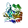
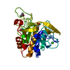
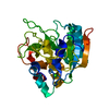
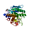
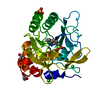
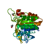
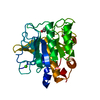
| Title | THE 0.78 ANGSTROMS STRUCTURE OF A SERINE PROTEASE-BACILLUS LENTUS SUBTILISIN | ||||||
 Components Components | SUBTILISIN | ||||||
 Keywords Keywords | SERINE PROTEASE / SUBTILISIN / BACILLUS LENTUS / HYDROLASE | ||||||
| Function / homology |  Function and homology information Function and homology informationsubtilisin / sporulation resulting in formation of a cellular spore / serine-type endopeptidase activity / proteolysis / extracellular region / metal ion binding Similarity search - Function | ||||||
| Biological species |  Bacillus lentus (bacteria) Bacillus lentus (bacteria) | ||||||
| Method |  X-RAY DIFFRACTION / SYNCHROTRON / MOLECULAR REPLACEMENT / Resolution: 0.78 Å X-RAY DIFFRACTION / SYNCHROTRON / MOLECULAR REPLACEMENT / Resolution: 0.78 Å | ||||||
 Authors Authors | Bott, R. / Kuhn, P. | ||||||
 Citation Citation | Journal: Biochemistry / Year: 1998 Title: The 0.78 A structure of a serine protease: Bacillus lentus subtilisin. Authors: Kuhn, P. / Knapp, M. / Soltis, S.M. / Ganshaw, G. / Thoene, M. / Bott, R. | ||||||
| History |
|
- Structure visualization
| Structure viewer | Molecule: MolmilJmol/JSmol |
|---|
- Downloads & links
-Download
| PDBx/mmCIF format | 1gci.cif.gz | 180.4 KB | Display | PDBx/mmCIF format |
|---|---|---|---|---|
| PDB format | pdb1gci.ent.gz | 138.7 KB | Display | PDB format |
| PDBx/mmJSON format | 1gci.json.gz | Tree view | PDBx/mmJSON format | |
| Others |  Other downloads Other downloads |
-Validation report
| Arichive directory | https://data.pdbj.org/pub/pdb/validation_reports/gc/1gciftp://data.pdbj.org/pub/pdb/validation_reports/gc/1gci | HTTPS FTP |
|---|
-Related structure data
| Related structure data | 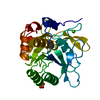 1jeaS S: Starting model for refinement |
|---|---|
| Similar structure data |


-Links
 PDBj
PDBj

- Assembly
| Deposited unit | 
| ||||||||
|---|---|---|---|---|---|---|---|---|---|
| 1 |
| ||||||||
| Unit cell |
|
-Components
| #1: Protein | Mass: 26718.381 Da / Num. of mol.: 1 Source method: isolated from a genetically manipulated source Source: (gene. exp.) Bacillus lentus (bacteria) / Production host: | ||||
|---|---|---|---|---|---|
| #2: Chemical | ChemComp-SO4 /   Mass: 96.063 Da / Num. of mol.: 1 / Source method: obtained synthetically / Formula: SO4 Mass: 96.063 Da / Num. of mol.: 1 / Source method: obtained synthetically / Formula: SO4 | ||||
| #3: Chemical |   Mass: 40.078 Da / Num. of mol.: 2 / Source method: obtained synthetically / Formula: Ca Mass: 40.078 Da / Num. of mol.: 2 / Source method: obtained synthetically / Formula: Ca#4: Chemical | ChemComp-GOL / |   Mass: 92.094 Da / Num. of mol.: 1 / Source method: obtained synthetically / Formula: C3H8O3 Mass: 92.094 Da / Num. of mol.: 1 / Source method: obtained synthetically / Formula: C3H8O3#5: Water | ChemComp-HOH / |  Mass: 18.015 Da / Num. of mol.: 384 / Source method: isolated from a natural source / Formula: H2O Mass: 18.015 Da / Num. of mol.: 384 / Source method: isolated from a natural source / Formula: H2O |
-Experimental details
-Experiment
| Experiment | Method: X-RAY DIFFRACTION / Number of used crystals: 1 |
|---|
- Sample preparation
| Crystal | Density Matthews: 2.31 Å3/Da / Density % sol: 47 % | ||||||||||||||||||||||||||||||||||||
|---|---|---|---|---|---|---|---|---|---|---|---|---|---|---|---|---|---|---|---|---|---|---|---|---|---|---|---|---|---|---|---|---|---|---|---|---|---|
| Crystal grow | pH: 5.9 / Details: FREE TEXT GOES HERE., pH 5.9 | ||||||||||||||||||||||||||||||||||||
| Crystal grow | *PLUS Temperature: 37 ℃ / Method: vapor diffusion, hanging drop | ||||||||||||||||||||||||||||||||||||
| Components of the solutions | *PLUS
|
-Data collection
| Diffraction | Mean temperature: 100 K |
|---|---|
| Diffraction source | Source: SYNCHROTRON / Site: SSRL  / Beamline: BL9-1 / Wavelength: 0.77 / Beamline: BL9-1 / Wavelength: 0.77 |
| Detector | Type: MAR scanner 300 mm plate / Detector: IMAGE PLATE / Date: Jun 10, 1997 / Details: MIRROR |
| Radiation | Monochromator: SI(111) / Monochromatic (M) / Laue (L): M / Scattering type: x-ray |
| Radiation wavelength | Wavelength: 0.77 Å / Relative weight: 1 |
| Reflection | Resolution: 0.78→35 Å / Num. obs: 257583 / % possible obs: 97.3 % / Observed criterion σ(I): 1 / Redundancy: 3.8 % / Rmerge(I) obs: 0.036 / Rsym value: 0.041 / Net I/σ(I): 5.8 |
| Reflection shell | Resolution: 0.78→0.82 Å / Redundancy: 3.7 % / Rmerge(I) obs: 0.29 / Mean I/σ(I) obs: 2.6 / Rsym value: 0.29 / % possible all: 92.7 |
| Reflection | *PLUS Num. measured all: 795681 |
| Reflection shell | *PLUS % possible obs: 92.7 % |
- Processing
| Software |
| |||||||||||||||||||||||||||||||||
|---|---|---|---|---|---|---|---|---|---|---|---|---|---|---|---|---|---|---|---|---|---|---|---|---|---|---|---|---|---|---|---|---|---|---|
| Refinement | Method to determine structure: MOLECULAR REPLACEMENT Starting model: 1JEA Resolution: 0.78→35 Å / Cross valid method: FREE R / σ(F): 0 Details: AMIDE HYDROGEN BOND LENGTHS IN THE PRESENT MODEL CLUSTER ABOUT 0.86 ANGSTROMS AND AROMATIC CARBON HYDROGEN DISTANCES CLUSTER ABOUT 0.96 ANGSTROMS. THESE DISTANCES SHOULD BE REGARDED AS ...Details: AMIDE HYDROGEN BOND LENGTHS IN THE PRESENT MODEL CLUSTER ABOUT 0.86 ANGSTROMS AND AROMATIC CARBON HYDROGEN DISTANCES CLUSTER ABOUT 0.96 ANGSTROMS. THESE DISTANCES SHOULD BE REGARDED AS PRELIMINARY RESULTS, PENDING FURTHER ANALYSIS. RESIDUE PRO 168 IS A CIS PROLINE. RESIDUE CA 276 CA IS A CALCIUM ++ ION RESIDUE CA 277 CA REFINED AS CALCIUM ++ ION HAS LOW OCCUPANCY AND IS PROBABLY ANOTHER CATION, POSSIBLY POTASSIUM RESIDUE SO4 278 IS A SULFATE ION. RESIDUE GOL 301 IS A GLYCEROL MOLECULE. A SPECIAL HYDROGEN BOND EXISTS BETWEEN ASP 32 OD2 AND HIS 64 ND1. THIS HYDROGEN ATOM IS SHARED BETWEEN THESE RESIDUES. THE POSITION IS ESTIMATED FROM THE PEAK FOR THIS ATOM IN A FO-FC DIFFERENCE ELECTRON DENSITY MAP. THIS HYDROGEN ATOM HAS BEEN DESIGNATED AS HD1 OF HIS 64 TO CONFORM TO THE PDB NOMENCLATURE.
| |||||||||||||||||||||||||||||||||
| Refine analyze | Num. disordered residues: 16 | |||||||||||||||||||||||||||||||||
| Refinement step | Cycle: LAST / Resolution: 0.78→35 Å
| |||||||||||||||||||||||||||||||||
| Refine LS restraints |
|
