 Movie
Movie Controller


+ Open data

- Basic information
| Entry | Database: PDB / ID: 1gan | |||||||||
|---|---|---|---|---|---|---|---|---|---|---|
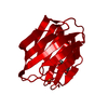
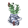
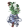
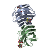
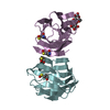
| Title | COMPLEX OF TOAD OVARY GALECTIN WITH N-ACETYLGALACTOSE | |||||||||
 Components Components | GALECTIN-1 | |||||||||
 Keywords Keywords | S-LECTIN / CARBOHYDRATE BINDING / LECTIN | |||||||||
| Function / homology |  Function and homology information Function and homology information | |||||||||
| Biological species | Bufo arenarum (Argentine toad) | |||||||||
| Method |  X-RAY DIFFRACTION / MOLECULAR REPLACEMENT / Resolution: 2.23 Å X-RAY DIFFRACTION / MOLECULAR REPLACEMENT / Resolution: 2.23 Å | |||||||||
 Authors Authors | Amzel, L.M. / Bianchet, M.A. / Ahmed, H. / Vasta, G.R. | |||||||||
 Citation Citation | Journal: Proteins / Year: 2000 Title: Soluble beta-galactosyl-binding lectin (galectin) from toad ovary: crystallographic studies of two protein-sugar complexes. Authors: Bianchet, M.A. / Ahmed, H. / Vasta, G.R. / Amzel, L.M. | |||||||||
| History |
|
- Structure visualization
| Structure viewer | Molecule: MolmilJmol/JSmol |
|---|
- Downloads & links
-Download
| PDBx/mmCIF format | 1gan.cif.gz | 65.3 KB | Display | PDBx/mmCIF format |
|---|---|---|---|---|
| PDB format | pdb1gan.ent.gz | 48.4 KB | Display | PDB format |
| PDBx/mmJSON format | 1gan.json.gz | Tree view | PDBx/mmJSON format | |
| Others |  Other downloads Other downloads |
-Validation report
| Arichive directory | https://data.pdbj.org/pub/pdb/validation_reports/ga/1ganftp://data.pdbj.org/pub/pdb/validation_reports/ga/1gan | HTTPS FTP |
|---|
-Related structure data
| Related structure data |  1a78C 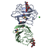 1sltS S: Starting model for refinement C: citing same article ( |
|---|---|
| Similar structure data |


-Links
 PDBj
PDBj- Assembly
| Deposited unit | 
| ||||||||
|---|---|---|---|---|---|---|---|---|---|
| 1 |
| ||||||||
| Unit cell |
| ||||||||
| Noncrystallographic symmetry (NCS) | NCS oper: (Code: given Matrix: (-0.810039, -0.423742, 0.405315), Vector: |
-Components
| #1: Protein | Mass: 14697.648 Da / Num. of mol.: 2 / Source method: isolated from a natural source / Source: (natural) Bufo arenarum (Argentine toad) / Organ: OVARY / References: UniProt: P56217 #2: Polysaccharide | 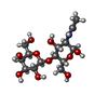  Source method: isolated from a genetically manipulated source Details: oligosaccharide / References: N-acetyl-alpha-lactosamine #3: Water | ChemComp-HOH / |  Mass: 18.015 Da / Num. of mol.: 30 / Source method: isolated from a natural source / Formula: H2O Mass: 18.015 Da / Num. of mol.: 30 / Source method: isolated from a natural source / Formula: H2O |
|---|
-Experimental details
-Experiment
| Experiment | Method: X-RAY DIFFRACTION / Number of used crystals: 1 |
|---|
- Sample preparation
| Crystal | Density Matthews: 2.34 Å3/Da / Density % sol: 47.45 % | ||||||||||||||||||||||||||||||||||||||||||
|---|---|---|---|---|---|---|---|---|---|---|---|---|---|---|---|---|---|---|---|---|---|---|---|---|---|---|---|---|---|---|---|---|---|---|---|---|---|---|---|---|---|---|---|
| Crystal grow | pH: 6.6 Details: DROPS OF EQUAL AMOUNT OF 10-12 MG/ML PROTEIN AND RESERVOIR SOLUTION WERE EQUILIBRATED AGAINST 1 ML OF (NH4)2SO4 AT 56% SATURATION IN 100MM TRIS-ACETATE BUFFER, PH 6.6 AND 1% MPD AND 1% DTT | ||||||||||||||||||||||||||||||||||||||||||
| Crystal grow | *PLUS Method: vapor diffusionDetails: drop consists of equal volume of protein and reservoir solutions | ||||||||||||||||||||||||||||||||||||||||||
| Components of the solutions | *PLUS
|
-Data collection
| Diffraction | Mean temperature: 300 K |
|---|---|
| Diffraction source | Source: ROTATING ANODE / Type: RIGAKU RUH3R / Wavelength: 1.5418 |
| Detector | Type: RIGAKU RAXIS / Detector: IMAGE PLATE / Date: Dec 1, 1994 / Details: MONOCHROMATOR |
| Radiation | Monochromator: GRAPHITE(002) / Monochromatic (M) / Laue (L): M / Scattering type: x-ray |
| Radiation wavelength | Wavelength: 1.5418 Å / Relative weight: 1 |
| Reflection | Resolution: 2.23→30 Å / Num. obs: 13192 / % possible obs: 90.9 % / Observed criterion σ(I): 0.5 / Redundancy: 7.5 % / Rmerge(I) obs: 0.075 / Rsym value: 0.075 / Net I/σ(I): 10000 |
| Reflection shell | Resolution: 2.25→2.5 Å / Redundancy: 7.49 % / Mean I/σ(I) obs: 3.3 / Rsym value: 0.19 / % possible all: 73.1 |
| Reflection | *PLUS Num. all: 13192 / Num. obs: 11873 / % possible obs: 901 % |
| Reflection shell | *PLUS % possible obs: 73.1 % / Rmerge(I) obs: 0.19 |
- Processing
| Software |
| ||||||||||||||||||||||||||||||||||||||||||||||||||||||||||||
|---|---|---|---|---|---|---|---|---|---|---|---|---|---|---|---|---|---|---|---|---|---|---|---|---|---|---|---|---|---|---|---|---|---|---|---|---|---|---|---|---|---|---|---|---|---|---|---|---|---|---|---|---|---|---|---|---|---|---|---|---|---|
| Refinement | Method to determine structure: MOLECULAR REPLACEMENT Starting model: PDB ENTRY 1SLT Resolution: 2.23→6 Å / σ(F): 2
| ||||||||||||||||||||||||||||||||||||||||||||||||||||||||||||
| Displacement parameters | Biso mean: 33.31 Å2 | ||||||||||||||||||||||||||||||||||||||||||||||||||||||||||||
| Refine analyze | Luzzati d res low obs: 6 Å | ||||||||||||||||||||||||||||||||||||||||||||||||||||||||||||
| Refinement step | Cycle: LAST / Resolution: 2.23→6 Å
| ||||||||||||||||||||||||||||||||||||||||||||||||||||||||||||
| Refine LS restraints |
| ||||||||||||||||||||||||||||||||||||||||||||||||||||||||||||
| LS refinement shell | Resolution: 2.23→2.33 Å
| ||||||||||||||||||||||||||||||||||||||||||||||||||||||||||||
| Xplor file |
| ||||||||||||||||||||||||||||||||||||||||||||||||||||||||||||
| Software | *PLUS Name: X-PLOR / Version: 3.1 / Classification: refinement | ||||||||||||||||||||||||||||||||||||||||||||||||||||||||||||
| Refinement | *PLUS Rfactor Rfree: 0.245 | ||||||||||||||||||||||||||||||||||||||||||||||||||||||||||||
| Solvent computation | *PLUS | ||||||||||||||||||||||||||||||||||||||||||||||||||||||||||||
| Displacement parameters | *PLUS | ||||||||||||||||||||||||||||||||||||||||||||||||||||||||||||
| Refine LS restraints | *PLUS
|
