 Movie
Movie Controller

[English] 日本語
 Yorodumi
- PDB-1g85: CRYSTAL STRUCTURE OF BOVINE ODORANT BINDING PROTEIN COMPLEXED WIT... -
+ Open data

- Basic information
| Entry | Database: PDB / ID: 1g85 | ||||||
|---|---|---|---|---|---|---|---|
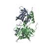
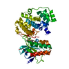
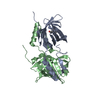
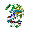
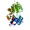
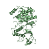
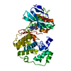
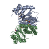
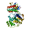
| Title | CRYSTAL STRUCTURE OF BOVINE ODORANT BINDING PROTEIN COMPLEXED WITH IS NATURAL LIGAND | ||||||
 Components Components | ODORANT-BINDING PROTEIN | ||||||
 Keywords Keywords | SIGNALING PROTEIN / lipocalin / swapping domain / homodimer | ||||||
| Function / homology |  Function and homology information Function and homology informationodorant binding / small molecule binding / sensory perception of smell / extracellular space Similarity search - Function | ||||||
| Biological species |  | ||||||
| Method |  X-RAY DIFFRACTION / MOLECULAR REPLACEMENT / Resolution: 1.8 Å X-RAY DIFFRACTION / MOLECULAR REPLACEMENT / Resolution: 1.8 Å | ||||||
 Authors Authors | Vincent, F. / Spinelli, S. / Cambillau, C. / Tegoni, M. | ||||||
 Citation Citation | Journal: J.Biol.Chem. / Year: 2001 Title: The insect attractant 1-octen-3-ol is the natural ligand of bovine odorant-binding protein. Authors: Ramoni, R. / Vincent, F. / Grolli, S. / Conti, V. / Malosse, C. / Boyer, F.D. / Nagnan-Le Meillour, P. / Spinelli, S. / Cambillau, C. / Tegoni, M. #1: Journal: Nat.Struct.Biol. / Year: 1996Title: Domain swapping creates a third putative combining site in bovine odorant binding protein dimer. Authors: Tegoni, M. / Spinelli, S. / Cambillau, C. / Spinelli, S. | ||||||
| History |
|
- Structure visualization
| Structure viewer | Molecule: MolmilJmol/JSmol |
|---|
- Downloads & links
-Download
| PDBx/mmCIF format | 1g85.cif.gz | 80.7 KB | Display | PDBx/mmCIF format |
|---|---|---|---|---|
| PDB format | pdb1g85.ent.gz | 61 KB | Display | PDB format |
| PDBx/mmJSON format | 1g85.json.gz | Tree view | PDBx/mmJSON format | |
| Others |  Other downloads Other downloads |
-Validation report
| Summary document | 1g85_validation.pdf.gz | 429 KB | Display | wwPDB validaton report |
|---|---|---|---|---|
| Full document | 1g85_full_validation.pdf.gz | 437.9 KB | Display | |
| Data in XML | 1g85_validation.xml.gz | 10.7 KB | Display | |
| Data in CIF | 1g85_validation.cif.gz | 14.9 KB | Display | |
| Arichive directory | https://data.pdbj.org/pub/pdb/validation_reports/g8/1g85ftp://data.pdbj.org/pub/pdb/validation_reports/g8/1g85 | HTTPS FTP |
-Related structure data
| Related structure data |  1hn2C 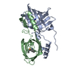 1pboS S: Starting model for refinement C: citing same article ( |
|---|---|
| Similar structure data |


-Links
 PDBj
PDBj

- Assembly
| Deposited unit | 
| ||||||||
|---|---|---|---|---|---|---|---|---|---|
| 1 |
| ||||||||
| Unit cell |
|
-Components
| #1: Protein | Mass: 18456.318 Da / Num. of mol.: 2 / Source method: isolated from a natural source / Source: (natural) #2: Chemical | ChemComp-3OL / (   Mass: 128.212 Da / Num. of mol.: 4 / Source method: obtained synthetically / Formula: C8H16O Mass: 128.212 Da / Num. of mol.: 4 / Source method: obtained synthetically / Formula: C8H16O#3: Water | ChemComp-HOH / |  Mass: 18.015 Da / Num. of mol.: 150 / Source method: isolated from a natural source / Formula: H2O Mass: 18.015 Da / Num. of mol.: 150 / Source method: isolated from a natural source / Formula: H2O |
|---|
-Experimental details
-Experiment
| Experiment | Method: X-RAY DIFFRACTION / Number of used crystals: 1 |
|---|
- Sample preparation
| Crystal | Density Matthews: 2.09 Å3/Da / Density % sol: 41.21 % |
|---|---|
| Crystal grow | Temperature: 277 K / Method: microdialysis / pH: 5.7 Details: 38% ethanol, 25mM sodium citrate, pH 5.7, MICRODIALYSIS, temperature 4K, temperature 277K |
-Data collection
| Diffraction | Mean temperature: 298 K |
|---|---|
| Diffraction source | Source: ROTATING ANODE / Type: RIGAKU RU200 / Wavelength: 1.5418 Å |
| Detector | Type: MARRESEARCH / Detector: IMAGE PLATE / Date: May 23, 2000 / Details: osmic mirrors |
| Radiation | Monochromator: osmic mirrors / Protocol: SINGLE WAVELENGTH / Monochromatic (M) / Laue (L): M / Scattering type: x-ray |
| Radiation wavelength | Wavelength: 1.5418 Å / Relative weight: 1 |
| Reflection | Resolution: 1.8→55 Å / Num. all: 237362 / Num. obs: 107952 / % possible obs: 45 % / Observed criterion σ(F): 2 / Observed criterion σ(I): 2 / Redundancy: 4 % / Biso Wilson estimate: 35.94 Å2 / Rmerge(I) obs: 0.084 / Net I/σ(I): 5 |
| Reflection shell | Resolution: 1.8→55 Å / Redundancy: 4 % / Rmerge(I) obs: 0.23 / Num. unique all: 27620 / % possible all: 97.8 |
- Processing
| Software |
| ||||||||||||||||||||
|---|---|---|---|---|---|---|---|---|---|---|---|---|---|---|---|---|---|---|---|---|---|
| Refinement | Method to determine structure: MOLECULAR REPLACEMENT Starting model: PDB entry 1PBO Resolution: 1.8→25.3 Å / Isotropic thermal model: RESTRAINED / Cross valid method: THROUGHOUT / σ(F): 2 / σ(I): 2 / Stereochemistry target values: Engh & Huber
| ||||||||||||||||||||
| Displacement parameters | Biso mean: 42.8 Å2
| ||||||||||||||||||||
| Refine analyze | Luzzati coordinate error obs: 0.21 Å / Luzzati d res low obs: 5 Å / Luzzati sigma a obs: 0.12 Å | ||||||||||||||||||||
| Refinement step | Cycle: LAST / Resolution: 1.8→25.3 Å
| ||||||||||||||||||||
| Refine LS restraints |
| ||||||||||||||||||||
| LS refinement shell | Resolution: 1.8→1.91 Å / Rfactor Rfree error: 0.02
|
