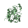
 Movie
Movie Controller


+ Open data

- Basic information
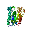
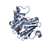
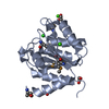
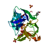
| Entry | Database: PDB / ID: 1g66 | |||||||||
|---|---|---|---|---|---|---|---|---|---|---|
| Title | ACETYLXYLAN ESTERASE AT 0.90 ANGSTROM RESOLUTION | |||||||||
 Components Components | ACETYL XYLAN ESTERASE II | |||||||||
 Keywords Keywords | HYDROLASE / serine hydrolase / acetyl xylopyranose / xylan | |||||||||
| Function / homology |  Function and homology information Function and homology informationacetylxylan esterase / acetylxylan esterase activity / xylan catabolic process / cellulose catabolic process / extracellular region Similarity search - Function | |||||||||
| Biological species |  Penicillium purpurogenum (fungus) Penicillium purpurogenum (fungus) | |||||||||
| Method |  X-RAY DIFFRACTION / SYNCHROTRON / AB INITIO PHASING / Resolution: 0.9 Å X-RAY DIFFRACTION / SYNCHROTRON / AB INITIO PHASING / Resolution: 0.9 Å | |||||||||
 Authors Authors | Ghosh, D. / Sawicki, M. / Lala, P. / Erman, M. / Pangborn, W. / Eyzaguirre, J. / Gutierrez, R. / Jornvall, H. / Thiel, D.J. | |||||||||
 Citation Citation | Journal: J.Biol.Chem. / Year: 2001 Title: Multiple conformations of catalytic serine and histidine in acetylxylan esterase at 0.90 A. Authors: Ghosh, D. / Sawicki, M. / Lala, P. / Erman, M. / Pangborn, W. / Eyzaguirre, J. / Gutierrez, R. / Jornvall, H. / Thiel, D.J. #1: Journal: Acta Crystallogr.,Sect.D / Year: 1999Title: Determination of a protein structure by iodination: the structure of iodinated acetylxylan esterase Authors: Ghosh, D. / Erman, M. / Sawicki, M. / Lala, P. / Weeks, D.R. / Li, N. / Pangborn, W. / Thiel, D.J. / Jornvall, H. / Gutierrez, R. / Eyzaguirre, J. #2: Journal: Proteins / Year: 1996Title: Characterization of crystals of P. purpurogenum acetyl xylan esterase from high resolution X-ray diffraction | |||||||||
| History |
|
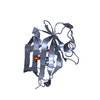
- Structure visualization
| Structure viewer | Molecule: MolmilJmol/JSmol |
|---|
- Downloads & links
-Download
| PDBx/mmCIF format | 1g66.cif.gz | 128.9 KB | Display | PDBx/mmCIF format |
|---|---|---|---|---|
| PDB format | pdb1g66.ent.gz | 102 KB | Display | PDB format |
| PDBx/mmJSON format | 1g66.json.gz | Tree view | PDBx/mmJSON format | |
| Others |  Other downloads Other downloads |
-Validation report
| Arichive directory | https://data.pdbj.org/pub/pdb/validation_reports/g6/1g66ftp://data.pdbj.org/pub/pdb/validation_reports/g6/1g66 | HTTPS FTP |
|---|
-Related structure data
| Related structure data | |
|---|---|
| Similar structure data |


-Links
 PDBj
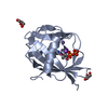
PDBj- Assembly
| Deposited unit | 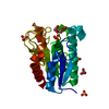
| ||||||||
|---|---|---|---|---|---|---|---|---|---|
| 1 |
| ||||||||
| Unit cell |
|
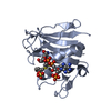
-Components
| #1: Protein | Mass: 20661.803 Da / Num. of mol.: 1 / Source method: isolated from a natural source / Source: (natural) Penicillium purpurogenum (fungus) / References: UniProt: O59893, acetylesterase | ||||||
|---|---|---|---|---|---|---|---|
| #2: Chemical | ChemComp-SO4 /   Mass: 96.063 Da / Num. of mol.: 4 / Source method: obtained synthetically / Formula: SO4 Mass: 96.063 Da / Num. of mol.: 4 / Source method: obtained synthetically / Formula: SO4#3: Chemical | ChemComp-GOL /   Mass: 92.094 Da / Num. of mol.: 4 / Source method: obtained synthetically / Formula: C3H8O3 Mass: 92.094 Da / Num. of mol.: 4 / Source method: obtained synthetically / Formula: C3H8O3#4: Water | ChemComp-HOH / |  Mass: 18.015 Da / Num. of mol.: 299 / Source method: isolated from a natural source / Formula: H2O Mass: 18.015 Da / Num. of mol.: 299 / Source method: isolated from a natural source / Formula: H2OHas protein modification | Y | |
-Experimental details
-Experiment
| Experiment | Method: X-RAY DIFFRACTION / Number of used crystals: 2 |
|---|
- Sample preparation
| Crystal | Density Matthews: 1.79 Å3/Da / Density % sol: 31.18 % | ||||||||||||||||||||||||||||||
|---|---|---|---|---|---|---|---|---|---|---|---|---|---|---|---|---|---|---|---|---|---|---|---|---|---|---|---|---|---|---|---|
| Crystal grow | Temperature: 298 K / Method: vapor diffusion / pH: 5.3 Details: Ammonium sulfate, pH 5.3, VAPOR DIFFUSION, temperature 298K | ||||||||||||||||||||||||||||||
| Crystal grow | *PLUS Method: vapor diffusion, hanging dropDetails: Pangborn, W., (1996) Proteons Struct.Funct.Genet., 24, 523. | ||||||||||||||||||||||||||||||
| Components of the solutions | *PLUS
|
-Data collection
| Diffraction | Mean temperature: 85 K |
|---|---|
| Diffraction source | Source: SYNCHROTRON / Site: CHESS  / Beamline: A1 / Beamline: A1 |
| Detector | Type: PRINCETON 2K / Detector: CCD / Date: Dec 15, 1995 |
| Radiation | Monochromator: Sagitally focussed Si (111) / Protocol: SINGLE WAVELENGTH / Monochromatic (M) / Laue (L): M / Scattering type: x-ray |
| Radiation wavelength | Relative weight: 1 |
| Reflection | Resolution: 0.9→99 Å / Num. all: 95283 / Num. obs: 95283 / % possible obs: 86.6 % / Observed criterion σ(F): 0 / Observed criterion σ(I): 0 / Redundancy: 4.41 % / Biso Wilson estimate: 10 Å2 / Rmerge(I) obs: 0.058 / Net I/σ(I): 29.2 |
| Reflection shell | Resolution: 0.9→0.94 Å / Redundancy: 2 % / Rmerge(I) obs: 0.185 / % possible all: 40.4 |
| Reflection | *PLUS Num. measured all: 420882 |
| Reflection shell | *PLUS % possible obs: 40.4 % |
- Processing
| Software |
| ||||||||||||||||||||
|---|---|---|---|---|---|---|---|---|---|---|---|---|---|---|---|---|---|---|---|---|---|
| Refinement | Method to determine structure: AB INITIO PHASING / Resolution: 0.9→99 Å / σ(F): 0 / σ(I): 0 / Stereochemistry target values: Engh & Huber
| ||||||||||||||||||||
| Refine analyze | Luzzati coordinate error obs: 0.1 Å | ||||||||||||||||||||
| Refinement step | Cycle: LAST / Resolution: 0.9→99 Å
| ||||||||||||||||||||
| Refine LS restraints |
| ||||||||||||||||||||
| Software | *PLUS Name: SHELXL-97 / Classification: refinement | ||||||||||||||||||||
| Refinement | *PLUS Rfactor obs: 0.103 | ||||||||||||||||||||
| Solvent computation | *PLUS | ||||||||||||||||||||
| Displacement parameters | *PLUS | ||||||||||||||||||||
| Refine LS restraints | *PLUS
|
