- PDB-2iqx: Rat Phosphatidylethanolamine-Binding Protein Containing the S153E... -
+
Open data
ID or keywords:
Loading...
-
Basic information
Entry
Database: PDB / ID: 2iqx
Title
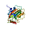
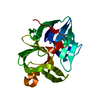
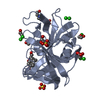
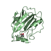
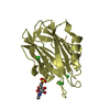
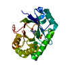
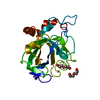
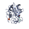
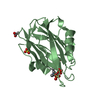
Rat Phosphatidylethanolamine-Binding Protein Containing the S153E Mutation in the Complex with o-Phosphorylethanolamine
Components
Phosphatidylethanolamine-binding protein 1
Keywords
HYDROLASE INHIBITOR / alpha-beta
Function / homology
Function and homology information
positive regulation of acetylcholine metabolic process / positive regulation of acetylcholine biosynthetic process / Negative regulation of MAPK pathway / MAP2K and MAPK activation / regulation of the force of heart contraction / receptor serine/threonine kinase binding / mitogen-activated protein kinase binding / sperm capacitation / response to corticosterone / eating behavior ...positive regulation of acetylcholine metabolic process / positive regulation of acetylcholine biosynthetic process / Negative regulation of MAPK pathway / MAP2K and MAPK activation / regulation of the force of heart contraction / receptor serine/threonine kinase binding / mitogen-activated protein kinase binding / sperm capacitation / response to corticosterone / eating behavior / spermatid development / negative regulation of protein phosphorylation / positive regulation of cAMP/PKA signal transduction / response to electrical stimulus / response to cAMP / negative regulation of MAPK cascade / positive regulation of mitotic nuclear division / axon terminus / response to activity / hippocampus development / serine-type endopeptidase inhibitor activity / response to calcium ion / response to toxic substance / kinase binding / apical part of cell / MAPK cascade / synaptic vesicle / response to oxidative stress / response to ethanol / mitochondrial outer membrane / response to xenobiotic stimulus / signaling receptor binding / neuronal cell body / lipid binding / protein kinase binding / enzyme binding / cell surface / : / ATP binding Similarity search - Function
Phosphatidylethanolamine-binding, conserved site / Phosphatidylethanolamine-binding protein family signature. / Phosphatidylethanolamine-binding Protein / PEBP-like / Phosphatidylethanolamine-binding protein, eukaryotic / Phosphatidylethanolamine-binding protein / Phosphatidylethanolamine-binding protein / PEBP-like superfamily / Alpha-Beta Complex / Alpha Beta Similarity search - Domain/homology
A: Phosphatidylethanolamine-binding protein 1 B: Phosphatidylethanolamine-binding protein 1 C: Phosphatidylethanolamine-binding protein 1 hetero molecules
In the structure databanks used in Yorodumi, some data are registered as the other names, "COVID-19 virus" and "2019-nCoV". Here are the details of the virus and the list of structure data.
Jan 31, 2019. EMDB accession codes are about to change! (news from PDBe EMDB page)
EMDB accession codes are about to change! (news from PDBe EMDB page)
The allocation of 4 digits for EMDB accession codes will soon come to an end. Whilst these codes will remain in use, new EMDB accession codes will include an additional digit and will expand incrementally as the available range of codes is exhausted. The current 4-digit format prefixed with “EMD-” (i.e. EMD-XXXX) will advance to a 5-digit format (i.e. EMD-XXXXX), and so on. It is currently estimated that the 4-digit codes will be depleted around Spring 2019, at which point the 5-digit format will come into force.
The EM Navigator/Yorodumi systems omit the EMD- prefix.
Related info.:Q: What is EMD? / ID/Accession-code notation in Yorodumi/EM Navigator
Yorodumi is a browser for structure data from EMDB, PDB, SASBDB, etc.
This page is also the successor to EM Navigator detail page, and also detail information page/front-end page for Omokage search.
The word "yorodu" (or yorozu) is an old Japanese word meaning "ten thousand". "mi" (miru) is to see.
Related info.:EMDB / PDB / SASBDB / Comparison of 3 databanks / Yorodumi Search / Aug 31, 2016. New EM Navigator & Yorodumi / Yorodumi Papers / Jmol/JSmol / Function and homology information / Changes in new EM Navigator and Yorodumi
 Movie
Movie Controller

 Yorodumi
 Open data
 Basic information
 Components
 Keywords
 Function and homology information
 X-RAY DIFFRACTION /
  Authors
 Citation
 Structure visualization
 Downloads & links
 Other downloads
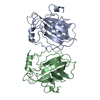


 PDBj
PDBj
 Assembly


 Mass: 141.063 Da / Num. of mol.: 3 / Source method: obtained synthetically / Formula: C2H8NO4P
 Mass: 18.015 Da / Num. of mol.: 573 / Source method: isolated from a natural source / Formula: H2O
 Sample preparation
 / Beamline: 19-ID / Wavelength: 0.97932
  Processing