 Movie
Movie Controller

[English] 日本語
 Yorodumi
- PDB-1fyu: Crystal structure of erythrina corallodendron lectin in hexagonal... -
+ Open data

- Basic information
| Entry | Database: PDB / ID: 1fyu | |||||||||
|---|---|---|---|---|---|---|---|---|---|---|
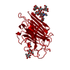
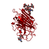
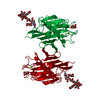
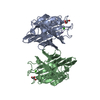
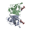
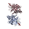
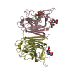
| Title | Crystal structure of erythrina corallodendron lectin in hexagonal crystal form | |||||||||
 Components Components | LECTIN | |||||||||
 Keywords Keywords | SUGAR BINDING PROTEIN / LECTIN / PROTEIN-CARBOHYDRATE COMPLEX | |||||||||
| Function / homology |  Function and homology information Function and homology information | |||||||||
| Biological species |  Erythrina corallodendron (coral tree) Erythrina corallodendron (coral tree) | |||||||||
| Method |  X-RAY DIFFRACTION / MOLECULAR REPLACEMEN / Resolution: 2.6 Å X-RAY DIFFRACTION / MOLECULAR REPLACEMEN / Resolution: 2.6 Å | |||||||||
 Authors Authors | Elgavish, S. / Shaanan, B. | |||||||||
 Citation Citation | Journal: Protein Sci. / Year: 2001 Title: Chemical characteristics of dimer interfaces in the legume lectin family. Authors: Elgavish, S. / Shaanan, B. #1: Journal: J.Mol.Biol. / Year: 1998Title: Structure of the Erythrina Corallodendron Lectin and of its Complexes with Mono- and Disaccharides | |||||||||
| History |
|
- Structure visualization
| Structure viewer | Molecule: MolmilJmol/JSmol |
|---|
- Downloads & links
-Download
| PDBx/mmCIF format | 1fyu.cif.gz | 105.6 KB | Display | PDBx/mmCIF format |
|---|---|---|---|---|
| PDB format | pdb1fyu.ent.gz | 80.8 KB | Display | PDB format |
| PDBx/mmJSON format | 1fyu.json.gz | Tree view | PDBx/mmJSON format | |
| Others |  Other downloads Other downloads |
-Validation report
| Arichive directory | https://data.pdbj.org/pub/pdb/validation_reports/fy/1fyuftp://data.pdbj.org/pub/pdb/validation_reports/fy/1fyu | HTTPS FTP |
|---|
-Related structure data
| Related structure data | 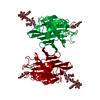 1lteS S: Starting model for refinement |
|---|---|
| Similar structure data |


-Links
 PDBj
PDBj

- Assembly
| Deposited unit | 
| ||||||||
|---|---|---|---|---|---|---|---|---|---|
| 1 |
| ||||||||
| Unit cell |
|
-Components
| #1: Protein | Mass: 27919.006 Da / Num. of mol.: 2 / Source method: isolated from a natural source / Source: (natural) Erythrina corallodendron (coral tree) / References: UniProt: P16404#2: Polysaccharide |   Source method: isolated from a genetically manipulated source Details: oligosaccharide / References: alpha-lactose #3: Chemical |   Mass: 54.938 Da / Num. of mol.: 2 / Source method: obtained synthetically / Formula: Mn Mass: 54.938 Da / Num. of mol.: 2 / Source method: obtained synthetically / Formula: Mn#4: Chemical |   Mass: 40.078 Da / Num. of mol.: 2 / Source method: obtained synthetically / Formula: Ca Mass: 40.078 Da / Num. of mol.: 2 / Source method: obtained synthetically / Formula: Ca#5: Water | ChemComp-HOH / |  Mass: 18.015 Da / Num. of mol.: 202 / Source method: isolated from a natural source / Formula: H2O Mass: 18.015 Da / Num. of mol.: 202 / Source method: isolated from a natural source / Formula: H2O |
|---|
-Experimental details
-Experiment
| Experiment | Method: X-RAY DIFFRACTION / Number of used crystals: 1 |
|---|
- Sample preparation
| Crystal | Density Matthews: 3.6 Å3/Da / Density % sol: 60 % | ||||||||||||||||||||||||||||||||||||||||
|---|---|---|---|---|---|---|---|---|---|---|---|---|---|---|---|---|---|---|---|---|---|---|---|---|---|---|---|---|---|---|---|---|---|---|---|---|---|---|---|---|---|
| Crystal grow | Temperature: 298 K / Method: vapor diffusion, hanging drop / pH: 7 Details: PEG 8000, sodium chloride, Hepes buffer, sodium azides, pH 7.0, VAPOR DIFFUSION, HANGING DROP, temperature 298K | ||||||||||||||||||||||||||||||||||||||||
| Crystal grow | *PLUS Details: Elgavish, S., (1998) J. Mol. Biol., 277, 917. | ||||||||||||||||||||||||||||||||||||||||
| Components of the solutions | *PLUS
|
-Data collection
| Diffraction | Mean temperature: 298 K |
|---|---|
| Diffraction source | Source: ROTATING ANODE / Type: RIGAKU RU300 / Wavelength: 1.5418 |
| Detector | Type: RIGAKU RAXIS II / Detector: IMAGE PLATE / Date: Sep 4, 1995 |
| Radiation | Protocol: SINGLE WAVELENGTH / Monochromatic (M) / Laue (L): M / Scattering type: x-ray |
| Radiation wavelength | Wavelength: 1.5418 Å / Relative weight: 1 |
| Reflection | Resolution: 2.6→39.16 Å / Num. obs: 26125 / % possible obs: 97.3 % / Redundancy: 8.5 % / Biso Wilson estimate: 43.4 Å2 / Rsym value: 0.074 / Net I/σ(I): 8.9 |
| Reflection shell | Resolution: 2.6→2.66 Å / Redundancy: 8.6 % / Rsym value: 0.34 / % possible all: 59.9 |
| Reflection | *PLUS Highest resolution: 2.6 Å / Lowest resolution: 39.16 Å / Redundancy: 8.5 % / Biso Wilson estimate: 43.4 Å2 |
| Reflection shell | *PLUS Redundancy: 8.6 % |
- Processing
| Software |
| ||||||||||||||||||||||||||||||||||||||||||||||||||||||||||||
|---|---|---|---|---|---|---|---|---|---|---|---|---|---|---|---|---|---|---|---|---|---|---|---|---|---|---|---|---|---|---|---|---|---|---|---|---|---|---|---|---|---|---|---|---|---|---|---|---|---|---|---|---|---|---|---|---|---|---|---|---|---|
| Refinement | Method to determine structure: MOLECULAR REPLACEMEN Starting model: 1LTE.PDB Resolution: 2.6→39.16 Å / Rfactor Rfree error: 0.005 / Isotropic thermal model: GROUP / Cross valid method: THROUGHOUT
| ||||||||||||||||||||||||||||||||||||||||||||||||||||||||||||
| Solvent computation | Solvent model: FLAT MODEL / Bsol: 40.25 Å2 / ksol: 0.293 e/Å3 | ||||||||||||||||||||||||||||||||||||||||||||||||||||||||||||
| Displacement parameters | Biso mean: 38.2 Å2
| ||||||||||||||||||||||||||||||||||||||||||||||||||||||||||||
| Refine analyze |
| ||||||||||||||||||||||||||||||||||||||||||||||||||||||||||||
| Refinement step | Cycle: LAST / Resolution: 2.6→39.16 Å
| ||||||||||||||||||||||||||||||||||||||||||||||||||||||||||||
| Refine LS restraints |
| ||||||||||||||||||||||||||||||||||||||||||||||||||||||||||||
| Refine LS restraints NCS | NCS model details: CONSTR | ||||||||||||||||||||||||||||||||||||||||||||||||||||||||||||
| LS refinement shell | Resolution: 2.6→2.66 Å / Rfactor Rfree error: 0.037 / Total num. of bins used: 15
| ||||||||||||||||||||||||||||||||||||||||||||||||||||||||||||
| Xplor file |
| ||||||||||||||||||||||||||||||||||||||||||||||||||||||||||||
| Software | *PLUS Name: CNS / Classification: refinement | ||||||||||||||||||||||||||||||||||||||||||||||||||||||||||||
| Refinement | *PLUS % reflection Rfree: 6.8 % | ||||||||||||||||||||||||||||||||||||||||||||||||||||||||||||
| Solvent computation | *PLUS | ||||||||||||||||||||||||||||||||||||||||||||||||||||||||||||
| Displacement parameters | *PLUS Biso mean: 38.2 Å2 | ||||||||||||||||||||||||||||||||||||||||||||||||||||||||||||
| Refine LS restraints | *PLUS
| ||||||||||||||||||||||||||||||||||||||||||||||||||||||||||||
| LS refinement shell | *PLUS Rfactor Rfree: 0.316 / % reflection Rfree: 6.9 % / Rfactor Rwork: 0.237 |
