 Movie
Movie Controller

[English] 日本語
 Yorodumi
- PDB-1fym: SERENDIPITOUS CRYSTAL STRUCTURE CONTAINING THE HEAT SHOCK TRANSCR... -
+ Open data

- Basic information
| Entry | Database: PDB / ID: 1fym | ||||||
|---|---|---|---|---|---|---|---|
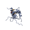
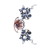
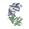
| Title | SERENDIPITOUS CRYSTAL STRUCTURE CONTAINING THE HEAT SHOCK TRANSCRIPTION FACTOR'S DNA BINDING DOMAIN AND COGNATE DNA IN A TAIL-TO-TAIL ORIENTATION | ||||||
 Components Components |
| ||||||
 Keywords Keywords | TRANSCRIPTION/DNA / crystal-packing interface / crystallization / protein-DNA interface / protein-protein interface / static disorder / TRANSCRIPTION-DNA COMPLEX | ||||||
| Function / homology |  Function and homology information Function and homology informationprotein-DNA complex / sequence-specific DNA binding / DNA-binding transcription factor activity / DNA binding / nucleus Similarity search - Function | ||||||
| Biological species |  Kluyveromyces lactis (yeast) Kluyveromyces lactis (yeast) | ||||||
| Method |  X-RAY DIFFRACTION / SYNCHROTRON / Resolution: 2.2 Å X-RAY DIFFRACTION / SYNCHROTRON / Resolution: 2.2 Å | ||||||
 Authors Authors | Littlefield, O. / Nelson, H.C.M. | ||||||
 Citation Citation | Journal: Proteins / Year: 2001 Title: Crystal packing interaction that blocks crystallization of a site-specific DNA binding protein-DNA complex. Authors: Littlefield, O. / Nelson, H.C. #1: Journal: Nat.Struct.Biol. / Year: 1999Title: A New Use for the 'Wing' of the 'Winged' Helix-Turn-Helix Motif in the HSF-DNA Cocrystal Authors: Littlefield, O. / Nelson, H.C.M. | ||||||
| History |
|
- Structure visualization
| Structure viewer | Molecule: MolmilJmol/JSmol |
|---|
- Downloads & links
-Download
| PDBx/mmCIF format | 1fym.cif.gz | 64.4 KB | Display | PDBx/mmCIF format |
|---|---|---|---|---|
| PDB format | pdb1fym.ent.gz | 44.2 KB | Display | PDB format |
| PDBx/mmJSON format | 1fym.json.gz | Tree view | PDBx/mmJSON format | |
| Others |  Other downloads Other downloads |
-Validation report
| Summary document | 1fym_validation.pdf.gz | 449.8 KB | Display | wwPDB validaton report |
|---|---|---|---|---|
| Full document | 1fym_full_validation.pdf.gz | 451.3 KB | Display | |
| Data in XML | 1fym_validation.xml.gz | 10.1 KB | Display | |
| Data in CIF | 1fym_validation.cif.gz | 13.4 KB | Display | |
| Arichive directory | https://data.pdbj.org/pub/pdb/validation_reports/fy/1fymftp://data.pdbj.org/pub/pdb/validation_reports/fy/1fym | HTTPS FTP |
-Related structure data


-Links
 PDBj
PDBj


- Assembly
| Deposited unit | 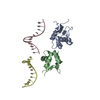
| ||||||||
|---|---|---|---|---|---|---|---|---|---|
| 1 | 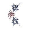
| ||||||||
| 2 | 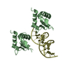
| ||||||||
| Unit cell |
| ||||||||
| Details | protein and DNA are not interacting in a physiologically relevant manner |
-Components
| #1: DNA chain | Mass: 3662.404 Da / Num. of mol.: 2 / Source method: obtained synthetically Details: This sequence is based on an idealized HSE sequence. #2: Protein | Mass: 10950.279 Da / Num. of mol.: 2 / Fragment: DNA BINDING DOMAIN / Mutation: N282R, F283H, K284A Source method: isolated from a genetically manipulated source Source: (gene. exp.) Kluyveromyces lactis (yeast) / Plasmid: PHN280R / Production host:  #3: Water | ChemComp-HOH / |  Mass: 18.015 Da / Num. of mol.: 92 / Source method: isolated from a natural source / Formula: H2O Mass: 18.015 Da / Num. of mol.: 92 / Source method: isolated from a natural source / Formula: H2O |
|---|
-Experimental details
-Experiment
| Experiment | Method: X-RAY DIFFRACTION / Number of used crystals: 1 |
|---|
- Sample preparation
| Crystal | Density Matthews: 2.09 Å3/Da / Density % sol: 41.23 % | ||||||||||||||||||||||||||||||||||||||||||||||||||||||||||||
|---|---|---|---|---|---|---|---|---|---|---|---|---|---|---|---|---|---|---|---|---|---|---|---|---|---|---|---|---|---|---|---|---|---|---|---|---|---|---|---|---|---|---|---|---|---|---|---|---|---|---|---|---|---|---|---|---|---|---|---|---|---|
| Crystal grow | Temperature: 277 K / Method: vapor diffusion, hanging drop / pH: 6 Details: PEG 4000, Isopropanol, Cacodylate, Ammonium Acetate, pH 6.0, VAPOR DIFFUSION, HANGING DROP, temperature 277K | ||||||||||||||||||||||||||||||||||||||||||||||||||||||||||||
| Components of the solutions |
| ||||||||||||||||||||||||||||||||||||||||||||||||||||||||||||
| Crystal grow | *PLUS Temperature: 4 ℃ / pH: 7.5 / Method: vapor diffusion | ||||||||||||||||||||||||||||||||||||||||||||||||||||||||||||
| Components of the solutions | *PLUS
|
-Data collection
| Diffraction | Mean temperature: 100 K |
|---|---|
| Diffraction source | Source: SYNCHROTRON / Site: SSRL  / Beamline: BL7-1 / Wavelength: 1.02 / Beamline: BL7-1 / Wavelength: 1.02 |
| Detector | Type: MARRESEARCH / Detector: IMAGE PLATE / Date: Jan 1, 1995 |
| Radiation | Protocol: SINGLE WAVELENGTH / Monochromatic (M) / Laue (L): M / Scattering type: x-ray |
| Radiation wavelength | Wavelength: 1.02 Å / Relative weight: 1 |
| Reflection | Resolution: 2.2→20 Å / Num. all: 10644 / Num. obs: 10644 / % possible obs: 84.5 % / Observed criterion σ(F): 0 / Observed criterion σ(I): 0 / Redundancy: 3.1 % / Rmerge(I) obs: 0.049 / Net I/σ(I): 12 |
| Reflection shell | Resolution: 2.2→2.25 Å / Redundancy: 2.8 % / Rmerge(I) obs: 0.186 / % possible all: 72.7 |
| Reflection | *PLUS |
| Reflection shell | *PLUS % possible obs: 72.7 % / Mean I/σ(I) obs: 4 |
- Processing
| Software |
| ||||||||||||||||||||
|---|---|---|---|---|---|---|---|---|---|---|---|---|---|---|---|---|---|---|---|---|---|
| Refinement | Resolution: 2.2→20 Å / σ(F): 0 / σ(I): 0 / Stereochemistry target values: Engh & Huber Details: The structure was originally refined by mistake with a glutamate at position 273. The deposited coordinates have been adjusted to contain aspartate at position 273, with the OD1 and OD2 ...Details: The structure was originally refined by mistake with a glutamate at position 273. The deposited coordinates have been adjusted to contain aspartate at position 273, with the OD1 and OD2 atoms having an arbitrary B-factor of 20.
| ||||||||||||||||||||
| Refinement step | Cycle: LAST / Resolution: 2.2→20 Å
| ||||||||||||||||||||
| Refine LS restraints |
| ||||||||||||||||||||
| Software | *PLUS Name: X-PLOR / Version: 3.1 / Classification: refinement | ||||||||||||||||||||
| Refinement | *PLUS Highest resolution: 2.2 Å / Lowest resolution: 20 Å / σ(F): 0 / Rfactor obs: 0.211 | ||||||||||||||||||||
| Solvent computation | *PLUS | ||||||||||||||||||||
| Displacement parameters | *PLUS | ||||||||||||||||||||
| LS refinement shell | *PLUS Highest resolution: 2.2 Å / Lowest resolution: 2.28 Å / Rfactor Rfree: 0.372 / Num. reflection Rfree: 77 / Num. reflection Rwork: 733 / Rfactor obs: 0.29 |
