 Movie
Movie Controller


+ Open data

- Basic information
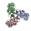
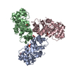
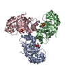
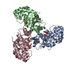
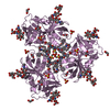
| Entry | Database: PDB / ID: 1fwr | ||||||
|---|---|---|---|---|---|---|---|
| Title | CRYSTAL STRUCTURE OF KDPG ALDOLASE DOUBLE MUTANT K133Q/T161K | ||||||
 Components Components | KDPG ALDOLASE | ||||||
 Keywords Keywords | LYASE / TIM barrel | ||||||
| Function / homology |  Function and homology information Function and homology information(4S)-4-hydroxy-2-oxoglutarate aldolase / (4S)-4-hydroxy-2-oxoglutarate aldolase activity / : / (R,S)-4-hydroxy-2-oxoglutarate aldolase activity / oxaloacetate decarboxylase / 2-dehydro-3-deoxy-phosphogluconate aldolase / oxo-acid-lyase activity / 2-dehydro-3-deoxy-phosphogluconate aldolase activity / oxaloacetate decarboxylase activity / aldehyde-lyase activity ...(4S)-4-hydroxy-2-oxoglutarate aldolase / (4S)-4-hydroxy-2-oxoglutarate aldolase activity / : / (R,S)-4-hydroxy-2-oxoglutarate aldolase activity / oxaloacetate decarboxylase / 2-dehydro-3-deoxy-phosphogluconate aldolase / oxo-acid-lyase activity / 2-dehydro-3-deoxy-phosphogluconate aldolase activity / oxaloacetate decarboxylase activity / aldehyde-lyase activity / membrane / identical protein binding / cytosol Similarity search - Function | ||||||
| Biological species |  | ||||||
| Method |  X-RAY DIFFRACTION / SYNCHROTRON / Resolution: 2.7 Å X-RAY DIFFRACTION / SYNCHROTRON / Resolution: 2.7 Å | ||||||
 Authors Authors | Naismith, J.H. / Buchanan, L.V. | ||||||
 Citation Citation | Journal: Structure / Year: 2001 Title: Directed evolution of a new catalytic site in 2-keto-3-deoxy-6-phosphogluconate aldolase from Escherichia coli. Authors: Wymer, N. / Buchanan, L.V. / Henderson, D. / Mehta, N. / Botting, C.H. / Pocivavsek, L. / Fierke, C.A. / Toone, E.J. / Naismith, J.H. | ||||||
| History |
|
- Structure visualization
| Structure viewer | Molecule: MolmilJmol/JSmol |
|---|
- Downloads & links
-Download
| PDBx/mmCIF format | 1fwr.cif.gz | 128.8 KB | Display | PDBx/mmCIF format |
|---|---|---|---|---|
| PDB format | pdb1fwr.ent.gz | 100.9 KB | Display | PDB format |
| PDBx/mmJSON format | 1fwr.json.gz | Tree view | PDBx/mmJSON format | |
| Others |  Other downloads Other downloads |
-Validation report
| Arichive directory | https://data.pdbj.org/pub/pdb/validation_reports/fw/1fwrftp://data.pdbj.org/pub/pdb/validation_reports/fw/1fwr | HTTPS FTP |
|---|
-Related structure data


-Links
 PDBj
PDBj- Assembly
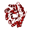
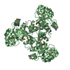
| Deposited unit | 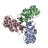
| ||||||||
|---|---|---|---|---|---|---|---|---|---|
| 1 |
| ||||||||
| Unit cell |
|
-Components
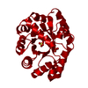
| #1: Protein | Mass: 22331.977 Da / Num. of mol.: 3 / Mutation: K133Q, T161K Source method: isolated from a genetically manipulated source Source: (gene. exp.) References: UniProt: P0A955, 2-dehydro-3-deoxy-phosphogluconate aldolase #2: Chemical |   Mass: 192.124 Da / Num. of mol.: 3 / Source method: obtained synthetically / Formula: C6H8O7 Mass: 192.124 Da / Num. of mol.: 3 / Source method: obtained synthetically / Formula: C6H8O7#3: Water | ChemComp-HOH / |  Mass: 18.015 Da / Num. of mol.: 137 / Source method: isolated from a natural source / Formula: H2O Mass: 18.015 Da / Num. of mol.: 137 / Source method: isolated from a natural source / Formula: H2O |
|---|
-Experimental details
-Experiment
| Experiment | Method: X-RAY DIFFRACTION / Number of used crystals: 1 |
|---|
- Sample preparation
| Crystal | Density Matthews: 2.33 Å3/Da / Density % sol: 47.31 % | ||||||||||||||||||||||||||||||
|---|---|---|---|---|---|---|---|---|---|---|---|---|---|---|---|---|---|---|---|---|---|---|---|---|---|---|---|---|---|---|---|
| Crystal grow | Temperature: 293 K / Method: vapor diffusion, hanging drop / pH: 4 Details: PEG 6K 20%, 0.075M citric acid, 30% sucrose, pH 4.0, VAPOR DIFFUSION, HANGING DROP, temperature 293K | ||||||||||||||||||||||||||||||
| Crystal grow | *PLUS pH: 8 / Method: vapor diffusion, sitting dropDetails: Buchanan, L.Vl., (1999) Acta Crystallogr., D55, 1946. | ||||||||||||||||||||||||||||||
| Components of the solutions | *PLUS
|
-Data collection
| Diffraction | Mean temperature: 100 K |
|---|---|
| Diffraction source | Source: SYNCHROTRON / Site: ESRF  / Beamline: ID14-1 / Wavelength: 0.934 / Beamline: ID14-1 / Wavelength: 0.934 |
| Detector | Type: ADSC QUANTUM 4 / Detector: CCD / Date: Jun 4, 2000 |
| Radiation | Protocol: SINGLE WAVELENGTH / Monochromatic (M) / Laue (L): M / Scattering type: x-ray |
| Radiation wavelength | Wavelength: 0.934 Å / Relative weight: 1 |
| Reflection | Resolution: 2.7→67 Å / Num. all: 57991 / Num. obs: 57991 / % possible obs: 99.9 % / Observed criterion σ(F): 2 / Observed criterion σ(I): 2 / Redundancy: 3.3 % / Biso Wilson estimate: 53.4 Å2 / Rmerge(I) obs: 0.087 / Net I/σ(I): 6.3 |
| Reflection shell | Resolution: 2.7→2.85 Å / Redundancy: 3.4 % / Rmerge(I) obs: 0.256 / Num. unique all: 8553 / % possible all: 99.8 |
| Reflection | *PLUS Lowest resolution: 67 Å / Num. obs: 17894 / % possible obs: 99.8 % |
| Reflection shell | *PLUS Highest resolution: 2.7 Å / % possible obs: 100 % / Mean I/σ(I) obs: 2.8 |
- Processing
| Software |
| ||||||||||||||||||||
|---|---|---|---|---|---|---|---|---|---|---|---|---|---|---|---|---|---|---|---|---|---|
| Refinement | Resolution: 2.7→67 Å / σ(F): 2 / σ(I): 2 / Stereochemistry target values: Engh & Huber
| ||||||||||||||||||||
| Refinement step | Cycle: LAST / Resolution: 2.7→67 Å
| ||||||||||||||||||||
| Refine LS restraints |
| ||||||||||||||||||||
| Software | *PLUS Name: CNS / Classification: refinement | ||||||||||||||||||||
| Refinement | *PLUS Highest resolution: 2.7 Å / Lowest resolution: 67 Å / σ(F): 2 / % reflection Rfree: 5 % | ||||||||||||||||||||
| Solvent computation | *PLUS | ||||||||||||||||||||
| Displacement parameters | *PLUS |
