 Movie
Movie Controller

[English] 日本語
 Yorodumi
- PDB-1eun: STRUCTURE OF 2-KETO-3-DEOXY-6-PHOSPHOGLUCONATE ALDOLASE FROM ESCH... -
+ Open data

- Basic information
| Entry | Database: PDB / ID: 1eun | ||||||
|---|---|---|---|---|---|---|---|
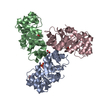
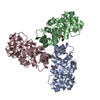
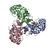
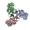
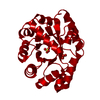
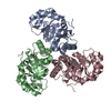
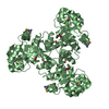
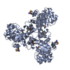
| Title | STRUCTURE OF 2-KETO-3-DEOXY-6-PHOSPHOGLUCONATE ALDOLASE FROM ESCHERICHIA COLI | ||||||
 Components Components | KDPG ALDOLASE | ||||||
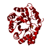
 Keywords Keywords | LYASE / 2-keto-3-deoxy-6-phosphogluconate aldolase / sulfate / trimer / beta-barrel | ||||||
| Function / homology |  Function and homology information Function and homology information(4S)-4-hydroxy-2-oxoglutarate aldolase / (4S)-4-hydroxy-2-oxoglutarate aldolase activity / : / (R,S)-4-hydroxy-2-oxoglutarate aldolase activity / oxaloacetate decarboxylase / 2-dehydro-3-deoxy-phosphogluconate aldolase / oxo-acid-lyase activity / 2-dehydro-3-deoxy-phosphogluconate aldolase activity / oxaloacetate decarboxylase activity / aldehyde-lyase activity ...(4S)-4-hydroxy-2-oxoglutarate aldolase / (4S)-4-hydroxy-2-oxoglutarate aldolase activity / : / (R,S)-4-hydroxy-2-oxoglutarate aldolase activity / oxaloacetate decarboxylase / 2-dehydro-3-deoxy-phosphogluconate aldolase / oxo-acid-lyase activity / 2-dehydro-3-deoxy-phosphogluconate aldolase activity / oxaloacetate decarboxylase activity / aldehyde-lyase activity / membrane / identical protein binding / cytosol Similarity search - Function | ||||||
| Biological species |  | ||||||
| Method |  X-RAY DIFFRACTION / SYNCHROTRON / Resolution: 2 Å X-RAY DIFFRACTION / SYNCHROTRON / Resolution: 2 Å | ||||||
 Authors Authors | Allard, J. / Grochulski, P. / Sygusch, J. | ||||||
 Citation Citation | Journal: Proc.Natl.Acad.Sci.USA / Year: 2001 Title: Covalent intermediate trapped in 2-keto-3-deoxy-6- phosphogluconate (KDPG) aldolase structure at 1.95-A resolution. Authors: Allard, J. / Grochulski, P. / Sygusch, J. | ||||||
| History |
|
- Structure visualization
| Structure viewer | Molecule: MolmilJmol/JSmol |
|---|
- Downloads & links
-Download
| PDBx/mmCIF format | 1eun.cif.gz | 144 KB | Display | PDBx/mmCIF format |
|---|---|---|---|---|
| PDB format | pdb1eun.ent.gz | 113.3 KB | Display | PDB format |
| PDBx/mmJSON format | 1eun.json.gz | Tree view | PDBx/mmJSON format | |
| Others |  Other downloads Other downloads |
-Validation report
| Arichive directory | https://data.pdbj.org/pub/pdb/validation_reports/eu/1eunftp://data.pdbj.org/pub/pdb/validation_reports/eu/1eun | HTTPS FTP |
|---|
-Related structure data


-Links
 PDBj
PDBj- Assembly
| Deposited unit | 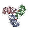
| ||||||||
|---|---|---|---|---|---|---|---|---|---|
| 1 |
| ||||||||
| Unit cell |
|
-Components
| #1: Protein | Mass: 22304.951 Da / Num. of mol.: 3 / Source method: isolated from a natural source / Source: (natural) References: UniProt: P0A955, 2-dehydro-3-deoxy-phosphogluconate aldolase #2: Chemical | ChemComp-SO4 /   Mass: 96.063 Da / Num. of mol.: 6 / Source method: obtained synthetically / Formula: SO4 Mass: 96.063 Da / Num. of mol.: 6 / Source method: obtained synthetically / Formula: SO4#3: Water | ChemComp-HOH / |  Mass: 18.015 Da / Num. of mol.: 749 / Source method: isolated from a natural source / Formula: H2O Mass: 18.015 Da / Num. of mol.: 749 / Source method: isolated from a natural source / Formula: H2O |
|---|
-Experimental details
-Experiment
| Experiment | Method: X-RAY DIFFRACTION / Number of used crystals: 1 |
|---|
- Sample preparation
| Crystal | Density Matthews: 2.34 Å3/Da / Density % sol: 47.37 % | ||||||||||||||||||||||||||||||
|---|---|---|---|---|---|---|---|---|---|---|---|---|---|---|---|---|---|---|---|---|---|---|---|---|---|---|---|---|---|---|---|
| Crystal grow | Temperature: 320 K / Method: vapor diffusion, hanging drop / pH: 4.6 Details: PEG 3350, Ammonium sulfate, Sodium Acetate, pH 4.6, VAPOR DIFFUSION, HANGING DROP, temperature 320K | ||||||||||||||||||||||||||||||
| Crystal grow | *PLUS | ||||||||||||||||||||||||||||||
| Components of the solutions | *PLUS
|
-Data collection
| Diffraction | Mean temperature: 180 K |
|---|---|
| Diffraction source | Source: SYNCHROTRON / Site: NSLS  / Beamline: X8C / Wavelength: 0.9789 / Beamline: X8C / Wavelength: 0.9789 |
| Detector | Type: ADSC QUANTUM 4r / Detector: CCD / Date: Oct 4, 1999 |
| Radiation | Protocol: SINGLE WAVELENGTH / Monochromatic (M) / Laue (L): M / Scattering type: x-ray |
| Radiation wavelength | Wavelength: 0.9789 Å / Relative weight: 1 |
| Reflection | Resolution: 1.8→40 Å / Num. all: 43245 / Num. obs: 39588 / % possible obs: 91.5 % / Observed criterion σ(F): 0 / Observed criterion σ(I): 0 / Redundancy: 4.66 % / Biso Wilson estimate: 12.6 Å2 / Rmerge(I) obs: 0.066 / Net I/σ(I): 20 |
| Reflection shell | Resolution: 2→2.13 Å / Redundancy: 4.38 % / Rmerge(I) obs: 0.269 / % possible all: 79.4 |
| Reflection | *PLUS Highest resolution: 2 Å / Num. obs: 39315 / % possible obs: 94.8 % / Rmerge(I) obs: 0.075 |
- Processing
| Software |
| ||||||||||||||||||||||||||||||||||||
|---|---|---|---|---|---|---|---|---|---|---|---|---|---|---|---|---|---|---|---|---|---|---|---|---|---|---|---|---|---|---|---|---|---|---|---|---|---|
| Refinement | Resolution: 2→39.49 Å / Rfactor Rfree error: 0.006 / Data cutoff high absF: 228160.38 / Data cutoff low absF: 0 / Isotropic thermal model: RESTRAINED / Cross valid method: THROUGHOUT / σ(F): 0 / σ(I): 0 / Stereochemistry target values: Engh & Huber
| ||||||||||||||||||||||||||||||||||||
| Solvent computation | Solvent model: FLAT MODEL / Bsol: 46.09 Å2 / ksol: 0.35 e/Å3 | ||||||||||||||||||||||||||||||||||||
| Displacement parameters | Biso mean: 28.3 Å2
| ||||||||||||||||||||||||||||||||||||
| Refine analyze |
| ||||||||||||||||||||||||||||||||||||
| Refinement step | Cycle: LAST / Resolution: 2→39.49 Å
| ||||||||||||||||||||||||||||||||||||
| Refine LS restraints |
| ||||||||||||||||||||||||||||||||||||
| LS refinement shell | Resolution: 2→2.13 Å / Rfactor Rfree error: 0.019 / Total num. of bins used: 6
| ||||||||||||||||||||||||||||||||||||
| Xplor file |
| ||||||||||||||||||||||||||||||||||||
| Software | *PLUS Name: CNS / Version: 1 / Classification: refinement | ||||||||||||||||||||||||||||||||||||
| Refine LS restraints | *PLUS
|
