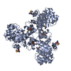
 Movie
Movie Controller

[English] 日本語
 Yorodumi
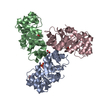
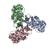
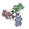
- PDB-1mxs: Crystal structure of 2-keto-3-deoxy-6-phosphogluconate (KDPG) ald... -
+ Open data

- Basic information
| Entry | Database: PDB / ID: 1mxs | ||||||
|---|---|---|---|---|---|---|---|
| Title | Crystal structure of 2-keto-3-deoxy-6-phosphogluconate (KDPG) aldolase from Pseudomonas putida. | ||||||
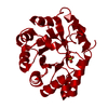
 Components Components | KDPG Aldolase | ||||||
 Keywords Keywords | LYASE / 2-KETO-3-DEOXY-6-PHOSPHOGLUCONATE ALDOLASE / SULFATE / BETA-BARREL | ||||||
| Function / homology |  Function and homology information Function and homology information2-dehydro-3-deoxy-phosphogluconate aldolase / 2-dehydro-3-deoxy-phosphogluconate aldolase activity Similarity search - Function | ||||||
| Biological species |  Pseudomonas putida (bacteria) Pseudomonas putida (bacteria) | ||||||
| Method |  X-RAY DIFFRACTION / MOLECULAR REPLACEMENT / Resolution: 2.2 Å X-RAY DIFFRACTION / MOLECULAR REPLACEMENT / Resolution: 2.2 Å | ||||||
 Authors Authors | Watanabe, L. / Bell, B.J. / Lebioda, L. / Rios-Steiner, J.L. / Tulinsky, A. / Arni, R.K. | ||||||
 Citation Citation | Journal: Acta Crystallogr.,Sect.D / Year: 2003 Title: Structure of 2-keto-3-deoxy-6-phosphogluconate (KDPG) aldolase from Pseudomonas putida. Authors: Bell, B.J. / Watanabe, L. / Rios-Steiner, J.L. / Tulinsky, A. / Lebioda, L. / Arni, R.K. #1: Journal: Structure / Year: 2001Title: Directed evolution of a new catalytic site in 2-keto-3-deoxy-6-phosphogluconate aldolase from Escherichia coli. Authors: Wymer, N. / Buchanan, L.V. / Henderson, D. / Mehta, N. / Botting, C.H. / Pocivavsek, L. / Fierke, C.A. / Toone, E.J. / Naismith, J.H. #2: Journal: Proc.Natl.Acad.Sci.USA / Year: 2001Title: Covalent intermediate trapped in 2-keto-3-deoxy-6-phosphogluconate (KDPG) aldolase structure at 1.95-A resolution Authors: Allard, J. / Grochulski, P. / Sygusch, J. #3: Journal: Biochemistry / Year: 1976Title: The folding and quaternary structure of trimeric 2-keto-3-deoxy-6-phosphogluconic aldolase at 3.5-A resolution. Authors: Mavridis, I.M. / Tulinsky, A. #4: Journal: J.Mol.Biol. / Year: 1982Title: Structure of 2-keto-3-deoxy-6-phosphogluconate aldolase at 2.8A resolution Authors: Mavridis, I.M. / Hatada, M.H. / Tulinsky, A. / Lebioda, L. | ||||||
| History |
|
- Structure visualization
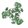
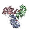
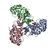
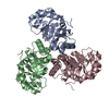
| Structure viewer | Molecule: MolmilJmol/JSmol |
|---|
- Downloads & links
-Download
| PDBx/mmCIF format | 1mxs.cif.gz | 55.6 KB | Display | PDBx/mmCIF format |
|---|---|---|---|---|
| PDB format | pdb1mxs.ent.gz | 39.8 KB | Display | PDB format |
| PDBx/mmJSON format | 1mxs.json.gz | Tree view | PDBx/mmJSON format | |
| Others |  Other downloads Other downloads |
-Validation report
| Arichive directory | https://data.pdbj.org/pub/pdb/validation_reports/mx/1mxsftp://data.pdbj.org/pub/pdb/validation_reports/mx/1mxs | HTTPS FTP |
|---|
-Related structure data
| Related structure data |  1eunS S: Starting model for refinement |
|---|---|
| Similar structure data |

-Links
 PDBj
PDBj- Assembly
| Deposited unit | 
| ||||||||
|---|---|---|---|---|---|---|---|---|---|
| 1 | 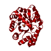
| ||||||||
| Unit cell |
| ||||||||
| Components on special symmetry positions |
|
-Components
| #1: Protein | Mass: 24003.762 Da / Num. of mol.: 1 / Source method: isolated from a natural source / Source: (natural) Pseudomonas putida (bacteria)References: UniProt: P00885, 2-dehydro-3-deoxy-phosphogluconate aldolase | ||
|---|---|---|---|
| #2: Chemical |   Mass: 96.063 Da / Num. of mol.: 2 / Source method: obtained synthetically / Formula: SO4 Mass: 96.063 Da / Num. of mol.: 2 / Source method: obtained synthetically / Formula: SO4#3: Water | ChemComp-HOH / |  Mass: 18.015 Da / Num. of mol.: 56 / Source method: isolated from a natural source / Formula: H2O Mass: 18.015 Da / Num. of mol.: 56 / Source method: isolated from a natural source / Formula: H2O |
-Experimental details
-Experiment
| Experiment | Method: X-RAY DIFFRACTION / Number of used crystals: 1 |
|---|
- Sample preparation
| Crystal | Density Matthews: 3.82 Å3/Da / Density % sol: 67.84 % | ||||||||||||||||||||||||||||
|---|---|---|---|---|---|---|---|---|---|---|---|---|---|---|---|---|---|---|---|---|---|---|---|---|---|---|---|---|---|
| Crystal grow | Temperature: 296 K / Method: microdialysis / pH: 3.5 Details: ammonium sulfate, KH2PO4, pH 3.5, MICRODIALYSIS, temperature 296K | ||||||||||||||||||||||||||||
| Crystal grow | *PLUS Method: microdialysis | ||||||||||||||||||||||||||||
| Components of the solutions | *PLUS
|
-Data collection
| Diffraction | Mean temperature: 296 K |
|---|---|
| Diffraction source | Source: ROTATING ANODE / Type: RIGAKU RU200 / Wavelength: 1.54 Å |
| Detector | Type: RIGAKU RAXIS II / Detector: IMAGE PLATE / Details: Yale/MSC mirrors |
| Radiation | Monochromator: GRAPHITE / Protocol: SINGLE WAVELENGTH / Monochromatic (M) / Laue (L): M / Scattering type: x-ray |
| Radiation wavelength | Wavelength: 1.54 Å / Relative weight: 1 |
| Reflection | Resolution: 2.2→9.98 Å / Num. obs: 9901 / Biso Wilson estimate: 12.6 Å2 / Rsym value: 0.046 |
| Reflection shell | Resolution: 2.2→50 Å / Num. unique all: 9901 / Rsym value: 0.046 |
| Reflection | *PLUS % possible obs: 52.3 % / Num. measured all: 66178 / Rmerge(I) obs: 0.046 |
- Processing
| Software |
| ||||||||||||||||||||
|---|---|---|---|---|---|---|---|---|---|---|---|---|---|---|---|---|---|---|---|---|---|
| Refinement | Method to determine structure: MOLECULAR REPLACEMENT Starting model: 1EUN Resolution: 2.2→9.98 Å / Rfactor Rfree error: 0.009 / Isotropic thermal model: RESTRAINED / Cross valid method: THROUGHOUT / σ(F): 3 / Stereochemistry target values: Engh & Huber
| ||||||||||||||||||||
| Solvent computation | Solvent model: FLAT MODEL / Bsol: 30.9183 Å2 / ksol: 0.341747 e/Å3 | ||||||||||||||||||||
| Displacement parameters | Biso mean: 34.9 Å2
| ||||||||||||||||||||
| Refine analyze |
| ||||||||||||||||||||
| Refinement step | Cycle: LAST / Resolution: 2.2→9.98 Å
| ||||||||||||||||||||
| Refine LS restraints |
| ||||||||||||||||||||
| LS refinement shell | Resolution: 2.2→50 Å / Rfactor Rfree error: 0.059 / Total num. of bins used: 6
| ||||||||||||||||||||
| Xplor file |
| ||||||||||||||||||||
| Refinement | *PLUS Highest resolution: 2.2 Å / Lowest resolution: 9999 Å / % reflection Rfree: 10 % | ||||||||||||||||||||
| Solvent computation | *PLUS | ||||||||||||||||||||
| Displacement parameters | *PLUS | ||||||||||||||||||||
| Refine LS restraints | *PLUS
|
