 Movie
Movie Controller


+ Open data

- Basic information
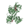
| Entry | Database: PDB / ID: 1fuj | |||||||||
|---|---|---|---|---|---|---|---|---|---|---|
| Title | PR3 (MYELOBLASTIN) | |||||||||
 Components Components | PR3 | |||||||||
 Keywords Keywords | HYDROLASE (SERINE PROTEASE) / HYDROLASE / SERINE PROTEASE / GLYCOPROTEIN / ZYMOGEN | |||||||||
| Function / homology |  Function and homology information Function and homology informationmyeloblastin / mature conventional dendritic cell differentiation / neutrophil extravasation / cell-cell junction maintenance / antimicrobial humoral response / Antimicrobial peptides / Other interleukin signaling / negative regulation of phagocytosis / collagen catabolic process / membrane protein ectodomain proteolysis ...myeloblastin / mature conventional dendritic cell differentiation / neutrophil extravasation / cell-cell junction maintenance / antimicrobial humoral response / Antimicrobial peptides / Other interleukin signaling / negative regulation of phagocytosis / collagen catabolic process / membrane protein ectodomain proteolysis / positive regulation of GTPase activity / plasma membrane raft / : / serine-type peptidase activity / azurophil granule lumen / signaling receptor binding / serine-type endopeptidase activity / positive regulation of cell population proliferation / Neutrophil degranulation / enzyme binding / proteolysis / : / extracellular exosome / extracellular region / plasma membrane / cytosol Similarity search - Function | |||||||||
| Biological species |  Homo sapiens (human) Homo sapiens (human) | |||||||||
| Method |  X-RAY DIFFRACTION / Resolution: 2.2 Å X-RAY DIFFRACTION / Resolution: 2.2 Å | |||||||||
 Authors Authors | Fujinaga, M. / Chernaia, M.M. / Halenbeck, R. / Koths, K. / James, M.N.G. | |||||||||
 Citation Citation | Journal: J.Mol.Biol. / Year: 1996 Title: The crystal structure of PR3, a neutrophil serine proteinase antigen of Wegener's granulomatosis antibodies. Authors: Fujinaga, M. / Chernaia, M.M. / Halenbeck, R. / Koths, K. / James, M.N. #1: Journal: Embo J. / Year: 1986Title: X-Ray Crystal Structure of the Complex of Human Leukocyte Elastase (Pmn Elastase) and the Third Domain of the Turkey Ovomucoid Inhibitor Authors: Bode, W. / Wei, A.Z. / Huber, R. / Meyer, E. / Travis, J. / Neumann, S. | |||||||||
| History |
|
- Structure visualization
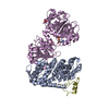
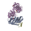
| Structure viewer | Molecule: MolmilJmol/JSmol |
|---|
- Downloads & links
-Download
| PDBx/mmCIF format | 1fuj.cif.gz | 184.3 KB | Display | PDBx/mmCIF format |
|---|---|---|---|---|
| PDB format | pdb1fuj.ent.gz | 147.5 KB | Display | PDB format |
| PDBx/mmJSON format | 1fuj.json.gz | Tree view | PDBx/mmJSON format | |
| Others |  Other downloads Other downloads |
-Validation report
| Arichive directory | https://data.pdbj.org/pub/pdb/validation_reports/fu/1fujftp://data.pdbj.org/pub/pdb/validation_reports/fu/1fuj | HTTPS FTP |
|---|
-Related structure data
| Similar structure data |
|---|


-Links
 PDBj
PDBj


- Assembly
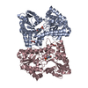
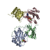
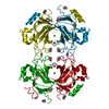
| Deposited unit | 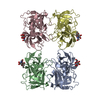
| ||||||||
|---|---|---|---|---|---|---|---|---|---|
| 1 |
| ||||||||
| Unit cell |
|
-Components
| #1: Protein | Mass: 24285.793 Da / Num. of mol.: 4 Source method: isolated from a genetically manipulated source Source: (gene. exp.) Homo sapiens (human) / Plasmid: BACULOVIRUS / Production host:  unidentified baculovirus / References: UniProt: P24158 unidentified baculovirus / References: UniProt: P24158#2: Polysaccharide | alpha-L-fucopyranose-(1-6)-2-acetamido-2-deoxy-beta-D-glucopyranose Source method: isolated from a genetically manipulated source #3: Water | ChemComp-HOH / |  Mass: 18.015 Da / Num. of mol.: 246 / Source method: isolated from a natural source / Formula: H2O Mass: 18.015 Da / Num. of mol.: 246 / Source method: isolated from a natural source / Formula: H2OHas protein modification | Y | Sequence details | RESIDUE NUMBERING IS ACCORDING TO ALIGNMENT WITH CHYMOTRYPS | |
|---|
-Experimental details
-Experiment
| Experiment | Method: X-RAY DIFFRACTION |
|---|
- Sample preparation
| Crystal | Density Matthews: 2.7 Å3/Da / Density % sol: 54.48 % | ||||||||||||||||||||||||||||||||||||||||||
|---|---|---|---|---|---|---|---|---|---|---|---|---|---|---|---|---|---|---|---|---|---|---|---|---|---|---|---|---|---|---|---|---|---|---|---|---|---|---|---|---|---|---|---|
| Crystal grow | *PLUS pH: 6.5 / Method: vapor diffusion, hanging drop / Details: used to seeding | ||||||||||||||||||||||||||||||||||||||||||
| Components of the solutions | *PLUS
|
-Data collection
| Radiation | Scattering type: x-ray |
|---|---|
| Radiation wavelength | Relative weight: 1 |
| Reflection | Resolution: 1.98→68.7 Å / Num. obs: 65602 / % possible obs: 90 % / Observed criterion σ(I): 0 |
| Reflection | *PLUS Num. measured all: 191704 / Rmerge(I) obs: 0.073 |
- Processing
| Software | Name: TNT / Classification: refinement | ||||||||||||||||||||||||||||||
|---|---|---|---|---|---|---|---|---|---|---|---|---|---|---|---|---|---|---|---|---|---|---|---|---|---|---|---|---|---|---|---|
| Refinement | Resolution: 2.2→10 Å / σ(F): 0 /
| ||||||||||||||||||||||||||||||
| Refinement step | Cycle: LAST / Resolution: 2.2→10 Å
| ||||||||||||||||||||||||||||||
| Refine LS restraints |
| ||||||||||||||||||||||||||||||
| Refinement | *PLUS Rfactor Rwork: 0.201 | ||||||||||||||||||||||||||||||
| Solvent computation | *PLUS | ||||||||||||||||||||||||||||||
| Displacement parameters | *PLUS | ||||||||||||||||||||||||||||||
| Refine LS restraints | *PLUS
|
