 Movie
Movie Controller

[English] 日本語
 Yorodumi
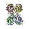
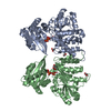
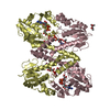
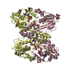
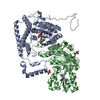
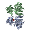
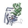
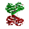
- PDB-1mfz: Partially refined 2.8 A Crystal structure of GDP-mannose dehydrog... -
+ Open data

- Basic information
| Entry | Database: PDB / ID: 1mfz | ||||||
|---|---|---|---|---|---|---|---|
| Title | Partially refined 2.8 A Crystal structure of GDP-mannose dehydrogenase from P. aeruginosa | ||||||
 Components Components | GDP-mannose 6-dehydrogenase | ||||||
 Keywords Keywords | OXIDOREDUCTASE / Rossmann fold / domain-swapped dimer / enzyme complex with cofactor and product | ||||||
| Function / homology |  Function and homology information Function and homology informationGDP-mannose 6-dehydrogenase / GDP-mannose 6-dehydrogenase activity / alginic acid biosynthetic process / single-species biofilm formation / response to osmotic stress / cellular response to cell envelope stress / NAD binding Similarity search - Function | ||||||
| Biological species |   Pseudomonas aeruginosa (bacteria) Pseudomonas aeruginosa (bacteria) | ||||||
| Method |  X-RAY DIFFRACTION / MOLECULAR REPLACEMENT / Resolution: 2.8 Å X-RAY DIFFRACTION / MOLECULAR REPLACEMENT / Resolution: 2.8 Å | ||||||
 Authors Authors | Snook, C.F. / Tipton, P.A. / Beamer, L.J. | ||||||
 Citation Citation | Journal: Biochemistry / Year: 2003 Title: The crystal structure of GDP-mannose dehydrogenase: A key enzyme in alginate biosynthesis of P. aeruginosa Authors: Snook, C.F. / Tipton, P.A. / Beamer, L.J. #1: Journal: Biochemistry / Year: 2002Title: Allosterism and cooperativity in Pseudomonas aeruginosa GDP-mannose dehydrogenase Authors: Naught, L.E. / Gilbert, S. / Imhoff, R. / Snook, C. / Beamer, L. / Tipton, P. | ||||||
| History |
|
- Structure visualization
| Structure viewer | Molecule: MolmilJmol/JSmol |
|---|
- Downloads & links
-Download
| PDBx/mmCIF format | 1mfz.cif.gz | 333 KB | Display | PDBx/mmCIF format |
|---|---|---|---|---|
| PDB format | pdb1mfz.ent.gz | 270.8 KB | Display | PDB format |
| PDBx/mmJSON format | 1mfz.json.gz | Tree view | PDBx/mmJSON format | |
| Others |  Other downloads Other downloads |
-Validation report
| Arichive directory | https://data.pdbj.org/pub/pdb/validation_reports/mf/1mfzftp://data.pdbj.org/pub/pdb/validation_reports/mf/1mfz | HTTPS FTP |
|---|
-Related structure data


-Links
 PDBj
PDBj


- Assembly
| Deposited unit | 
| ||||||||
|---|---|---|---|---|---|---|---|---|---|
| 1 | 
| ||||||||
| 2 | 
| ||||||||
| Unit cell |
|
-Components
| #1: Protein | Mass: 48078.469 Da / Num. of mol.: 4 Source method: isolated from a genetically manipulated source Source: (gene. exp.) Pseudomonas aeruginosa (bacteria) / Gene: AlgD / Plasmid: pET-3a / Species (production host): Escherichia coli / Production host: #2: Chemical | ChemComp-GDX /   Mass: 619.325 Da / Num. of mol.: 4 / Source method: obtained synthetically / Formula: C16H23N5O17P2 Mass: 619.325 Da / Num. of mol.: 4 / Source method: obtained synthetically / Formula: C16H23N5O17P2#3: Water | ChemComp-HOH / |  Mass: 18.015 Da / Num. of mol.: 106 / Source method: isolated from a natural source / Formula: H2O Mass: 18.015 Da / Num. of mol.: 106 / Source method: isolated from a natural source / Formula: H2OHas protein modification | Y | |
|---|
-Experimental details
-Experiment
| Experiment | Method: X-RAY DIFFRACTION / Number of used crystals: 1 |
|---|
- Sample preparation
| Crystal | Density Matthews: 2.55 Å3/Da / Density % sol: 51.74 % | ||||||||||||||||||||||||||||||||||||||||||
|---|---|---|---|---|---|---|---|---|---|---|---|---|---|---|---|---|---|---|---|---|---|---|---|---|---|---|---|---|---|---|---|---|---|---|---|---|---|---|---|---|---|---|---|
| Crystal grow | Method: vapor diffusion, hanging drop / pH: 5.6 Details: 10% PEG 3350, 20% isopropanol, 0.1 M Na citrate, pH 5.6, VAPOR DIFFUSION, HANGING DROP | ||||||||||||||||||||||||||||||||||||||||||
| Crystal grow | *PLUS pH: 7.5 | ||||||||||||||||||||||||||||||||||||||||||
| Components of the solutions | *PLUS
|
-Data collection
| Diffraction | Mean temperature: 100 K |
|---|---|
| Diffraction source | Source: ROTATING ANODE / Type: RIGAKU RU200 / Wavelength: 1.5418 |
| Detector | Type: RIGAKU RAXIS IV / Detector: IMAGE PLATE / Details: Osmic confocal |
| Radiation | Monochromator: Osmic confocal / Protocol: SINGLE WAVELENGTH / Monochromatic (M) / Laue (L): M / Scattering type: x-ray |
| Radiation wavelength | Wavelength: 1.5418 Å / Relative weight: 1 |
| Reflection | Resolution: 2.8→94.691 Å / Num. obs: 48049 / % possible obs: 98.7 % / Observed criterion σ(F): 0 / Observed criterion σ(I): 0 / Redundancy: 3.2 % / Biso Wilson estimate: 34.7 Å2 / Rmerge(I) obs: 0.065 / Net I/σ(I): 16.7 |
| Reflection shell | Resolution: 2.8→2.9 Å / Rmerge(I) obs: 0.217 / Mean I/σ(I) obs: 4.4 / Num. unique all: 4322 / % possible all: 90 |
| Reflection | *PLUS Num. measured all: 151736 |
| Reflection shell | *PLUS % possible obs: 90 % |
- Processing
| Software |
| |||||||||||||||||||||||||
|---|---|---|---|---|---|---|---|---|---|---|---|---|---|---|---|---|---|---|---|---|---|---|---|---|---|---|
| Refinement | Method to determine structure: MOLECULAR REPLACEMENT / Resolution: 2.8→40 Å / SU B: 14.6806 / SU ML: 0.30112 / Isotropic thermal model: isotropic / Cross valid method: THROUGHOUT / σ(F): 0 / ESU R Free: 0.40486 / Stereochemistry target values: Engh & Huber
| |||||||||||||||||||||||||
| Solvent computation | Ion probe radii: 0.8 Å / Shrinkage radii: 0.8 Å / VDW probe radii: 1.4 Å | |||||||||||||||||||||||||
| Displacement parameters | Biso mean: 34.708 Å2
| |||||||||||||||||||||||||
| Refinement step | Cycle: LAST / Resolution: 2.8→40 Å
| |||||||||||||||||||||||||
| Refine LS restraints |
| |||||||||||||||||||||||||
| LS refinement shell | Resolution: 2.8→2.9 Å / Rfactor Rfree error: 0.35
| |||||||||||||||||||||||||
| Refinement | *PLUS Lowest resolution: 40 Å / % reflection Rfree: 5 % / Rfactor Rfree: 0.263 / Rfactor Rwork: 0.207 | |||||||||||||||||||||||||
| Solvent computation | *PLUS | |||||||||||||||||||||||||
| Displacement parameters | *PLUS | |||||||||||||||||||||||||
| Refine LS restraints | *PLUS
|
