 Movie
Movie Controller

[English] 日本語
 Yorodumi
- PDB-1ft5: CRYSTAL STRUCTURE OF THE OXIDIZED STATE OF CYTOCHROME C554 FROM N... -
+ Open data

- Basic information
| Entry | Database: PDB / ID: 1ft5 | ||||||
|---|---|---|---|---|---|---|---|
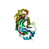
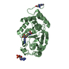
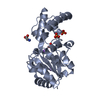
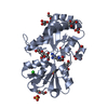
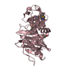
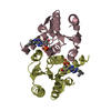
| Title | CRYSTAL STRUCTURE OF THE OXIDIZED STATE OF CYTOCHROME C554 FROM NITROSOMONAS EUROPAEA | ||||||
 Components Components | CYTOCHROME C554 | ||||||
 Keywords Keywords | ELECTRON TRANSPORT / heme-stacking | ||||||
| Function / homology |  Function and homology information Function and homology information | ||||||
| Biological species |  Nitrosomonas europaea (bacteria) Nitrosomonas europaea (bacteria) | ||||||
| Method |  X-RAY DIFFRACTION / SYNCHROTRON / Resolution: 1.6 Å X-RAY DIFFRACTION / SYNCHROTRON / Resolution: 1.6 Å | ||||||
 Authors Authors | Iverson, T.M. / Arciero, D.M. / Hooper, A.B. / Rees, D.C. | ||||||
 Citation Citation | Journal: J.Biol.Inorg.Chem. / Year: 2001 Title: High-resolution structures of the oxidized and reduced states of cytochrome c554 from Nitrosomonas europaea. Authors: Iverson, T.M. / Arciero, D.M. / Hooper, A.B. / Rees, D.C. #1: Journal: Nat.Struct.Biol. / Year: 1998Title: Heme Packing Motifs Revealed by the Crystal Structure of the Tetra-heme Cytochrome c554 from Nitrosomonas europaea Authors: Iverson, T.M. / Arciero, D.M. / Hsu, B.T. / Logan, M.S. / Hooper, A.B. / Rees, D.C. | ||||||
| History |
|
- Structure visualization
| Structure viewer | Molecule: MolmilJmol/JSmol |
|---|
- Downloads & links
-Download
| PDBx/mmCIF format | 1ft5.cif.gz | 64 KB | Display | PDBx/mmCIF format |
|---|---|---|---|---|
| PDB format | pdb1ft5.ent.gz | 47.3 KB | Display | PDB format |
| PDBx/mmJSON format | 1ft5.json.gz | Tree view | PDBx/mmJSON format | |
| Others |  Other downloads Other downloads |
-Validation report
| Arichive directory | https://data.pdbj.org/pub/pdb/validation_reports/ft/1ft5ftp://data.pdbj.org/pub/pdb/validation_reports/ft/1ft5 | HTTPS FTP |
|---|
-Related structure data


-Links
 PDBj
PDBj
- Assembly
| Deposited unit | 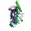
| ||||||||
|---|---|---|---|---|---|---|---|---|---|
| 1 |
| ||||||||
| Unit cell |
|
-Components
| #1: Protein | Mass: 23656.900 Da / Num. of mol.: 1 / Source method: isolated from a natural source Details: CHEMOAUTOTROPHIC BACTERIUM INVOLVED IN BIOLOGICAL NITRIFICATION Source: (natural) Nitrosomonas europaea (bacteria) / References: UniProt: Q57142 | ||||||
|---|---|---|---|---|---|---|---|
| #2: Chemical |   Mass: 94.971 Da / Num. of mol.: 3 / Source method: obtained synthetically / Formula: PO4 Mass: 94.971 Da / Num. of mol.: 3 / Source method: obtained synthetically / Formula: PO4#3: Chemical | ChemComp-HEM /   Mass: 616.487 Da / Num. of mol.: 4 / Source method: obtained synthetically / Formula: C34H32FeN4O4 Mass: 616.487 Da / Num. of mol.: 4 / Source method: obtained synthetically / Formula: C34H32FeN4O4#4: Water | ChemComp-HOH / |  Mass: 18.015 Da / Num. of mol.: 142 / Source method: isolated from a natural source / Formula: H2O Mass: 18.015 Da / Num. of mol.: 142 / Source method: isolated from a natural source / Formula: H2OHas protein modification | Y | |
-Experimental details
-Experiment
| Experiment | Method: X-RAY DIFFRACTION / Number of used crystals: 1 |
|---|
- Sample preparation
| Crystal | Density Matthews: 3.02 Å3/Da / Density % sol: 59.21 % | ||||||||||||||||||||||||
|---|---|---|---|---|---|---|---|---|---|---|---|---|---|---|---|---|---|---|---|---|---|---|---|---|---|
| Crystal grow | Temperature: 295 K / Method: vapor diffusion, sitting drop / pH: 10.1 Details: 62.5% w/vol potassium phosphate pH 10.1, VAPOR DIFFUSION, SITTING DROP, temperature 295K | ||||||||||||||||||||||||
| Crystal grow | *PLUS Temperature: 22 ℃ / Details: Iverson, T.M., (1998) Nat.Struct.Biol., 5, 1005. | ||||||||||||||||||||||||
| Components of the solutions | *PLUS
|
-Data collection
| Diffraction | Mean temperature: 93 K |
|---|---|
| Diffraction source | Source: SYNCHROTRON / Site: SSRL  / Beamline: BL7-1 / Wavelength: 1.08 / Beamline: BL7-1 / Wavelength: 1.08 |
| Detector | Type: MARRESEARCH / Detector: IMAGE PLATE / Date: Apr 23, 1998 |
| Radiation | Protocol: SINGLE WAVELENGTH / Monochromatic (M) / Laue (L): M / Scattering type: x-ray |
| Radiation wavelength | Wavelength: 1.08 Å / Relative weight: 1 |
| Reflection | Resolution: 1.6→20 Å / Num. all: 109601 / Num. obs: 35562 / % possible obs: 97.3 % / Observed criterion σ(F): 0 / Observed criterion σ(I): 0 / Redundancy: 3.08 % / Biso Wilson estimate: 18.6 Å2 / Rmerge(I) obs: 0.045 / Net I/σ(I): 28 |
| Reflection shell | Resolution: 1.6→1.68 Å / Rmerge(I) obs: 0.174 / % possible all: 86.1 |
| Reflection | *PLUS Num. measured all: 109601 |
| Reflection shell | *PLUS % possible obs: 86.1 % / Mean I/σ(I) obs: 7.3 |
- Processing
| Software |
| ||||||||||||||||||||
|---|---|---|---|---|---|---|---|---|---|---|---|---|---|---|---|---|---|---|---|---|---|
| Refinement | Resolution: 1.6→20 Å / σ(F): 0 / σ(I): 0 / Stereochemistry target values: Engh and Huber
| ||||||||||||||||||||
| Refinement step | Cycle: LAST / Resolution: 1.6→20 Å
| ||||||||||||||||||||
| Refine LS restraints |
| ||||||||||||||||||||
| Software | *PLUS Name: REFMAC / Classification: refinement | ||||||||||||||||||||
| Refinement | *PLUS Lowest resolution: 20 Å / σ(F): 0 / Rfactor obs: 0.188 | ||||||||||||||||||||
| Solvent computation | *PLUS | ||||||||||||||||||||
| Displacement parameters | *PLUS | ||||||||||||||||||||
| Refine LS restraints | *PLUS
|
