 Movie
Movie Controller

[English] 日本語
 Yorodumi
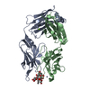
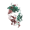
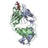
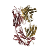
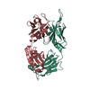
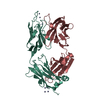
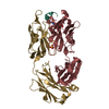
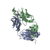
- PDB-1fpt: THREE-DIMENSIONAL STRUCTURE OF THE COMPLEX BETWEEN THE FAB FRAGME... -
+ Open data

- Basic information
| Entry | Database: PDB / ID: 1fpt | ||||||
|---|---|---|---|---|---|---|---|
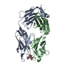
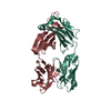
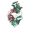
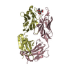
| Title | THREE-DIMENSIONAL STRUCTURE OF THE COMPLEX BETWEEN THE FAB FRAGMENT OF AN NEUTRALIZING ANTIBODY FOR TYPE 1 POLIOVIRUS AND ITS VIRAL EPITOPE | ||||||
 Components Components |
| ||||||
 Keywords Keywords | COMPLEX (ANTIBODY/PV-1 FRAGMENT) / COMPLEX (ANTIBODY-PV-1 FRAGMENT) / COMPLEX (ANTIBODY-PV-1 FRAGMENT) complex | ||||||
| Function / homology |  Function and homology information Function and homology informationpositive regulation of B cell activation / phagocytosis, recognition / humoral immune response mediated by circulating immunoglobulin / symbiont-mediated suppression of host translation initiation / early endosome to late endosome transport / positive regulation of type IIa hypersensitivity / positive regulation of type I hypersensitivity / antibody-dependent cellular cytotoxicity / Fc-gamma receptor I complex binding / immunoglobulin complex, circulating ...positive regulation of B cell activation / phagocytosis, recognition / humoral immune response mediated by circulating immunoglobulin / symbiont-mediated suppression of host translation initiation / early endosome to late endosome transport / positive regulation of type IIa hypersensitivity / positive regulation of type I hypersensitivity / antibody-dependent cellular cytotoxicity / Fc-gamma receptor I complex binding / immunoglobulin complex, circulating / phagocytosis, engulfment / immunoglobulin receptor binding / IgG immunoglobulin complex / antigen processing and presentation / endosome to lysosome transport / immunoglobulin mediated immune response / symbiont-mediated suppression of host cytoplasmic pattern recognition receptor signaling pathway via inhibition of RIG-I activity / regulation of proteolysis / complement activation, classical pathway / positive regulation of endocytosis / antigen binding / symbiont-mediated suppression of host cytoplasmic pattern recognition receptor signaling pathway via inhibition of MDA-5 activity / multivesicular body / symbiont-mediated suppression of host cytoplasmic pattern recognition receptor signaling pathway via inhibition of MAVS activity / positive regulation of phagocytosis / response to bacterium / picornain 2A / symbiont-mediated suppression of host mRNA export from nucleus / symbiont genome entry into host cell via pore formation in plasma membrane / picornain 3C / T=pseudo3 icosahedral viral capsid / host cell cytoplasmic vesicle membrane / positive regulation of immune response / ribonucleoside triphosphate phosphatase activity / nucleoside-triphosphate phosphatase / antibacterial humoral response / channel activity / monoatomic ion transmembrane transport / RNA helicase activity / endocytosis involved in viral entry into host cell / symbiont-mediated activation of host autophagy / RNA-directed RNA polymerase / cysteine-type endopeptidase activity / viral RNA genome replication / RNA-directed RNA polymerase activity / virion attachment to host cell / host cell nucleus / DNA-templated transcription / structural molecule activity / proteolysis / : / RNA binding / zinc ion binding / ATP binding / plasma membrane Similarity search - Function | ||||||
| Biological species |  Human poliovirus 1 Human poliovirus 1 | ||||||
| Method |  X-RAY DIFFRACTION / Resolution: 3 Å X-RAY DIFFRACTION / Resolution: 3 Å | ||||||
 Authors Authors | Wien, M.W. / Hogle, J.M. | ||||||
 Citation Citation | Journal: Nat.Struct.Biol. / Year: 1995 Title: Structure of the complex between the Fab fragment of a neutralizing antibody for type 1 poliovirus and its viral epitope. Authors: Wien, M.W. / Filman, D.J. / Stura, E.A. / Guillot, S. / Delpeyroux, F. / Crainic, R. / Hogle, J.M. | ||||||
| History |
|
- Structure visualization
| Structure viewer | Molecule: MolmilJmol/JSmol |
|---|
- Downloads & links
-Download
| PDBx/mmCIF format | 1fpt.cif.gz | 98.5 KB | Display | PDBx/mmCIF format |
|---|---|---|---|---|
| PDB format | pdb1fpt.ent.gz | 75.4 KB | Display | PDB format |
| PDBx/mmJSON format | 1fpt.json.gz | Tree view | PDBx/mmJSON format | |
| Others |  Other downloads Other downloads |
-Validation report
| Arichive directory | https://data.pdbj.org/pub/pdb/validation_reports/fp/1fptftp://data.pdbj.org/pub/pdb/validation_reports/fp/1fpt | HTTPS FTP |
|---|
-Related structure data
| Similar structure data |
|---|


-Links
 PDBj
PDBj


- Assembly
| Deposited unit | 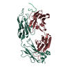
| ||||||||
|---|---|---|---|---|---|---|---|---|---|
| 1 |
| ||||||||
| Unit cell |
| ||||||||
| Atom site foot note | 1: CIS PROLINE - PRO L 8 / 2: CIS PROLINE - PRO L 95 / 3: CIS PROLINE - PRO L 141 4: ARG H 40 - PRO H 41 OMEGA = 247.33 PEPTIDE BOND DEVIATES SIGNIFICANTLY FROM TRANS CONFORMATION 5: CIS PROLINE - PRO H 149 / 6: CIS PROLINE - PRO H 151 |
-Components
| #1: Antibody | Mass: 1940.200 Da / Num. of mol.: 1 Source method: isolated from a genetically manipulated source Source: (gene. exp.) Human poliovirus 1 / Genus: Enterovirus / Species: Poliovirus / Strain: Mahoney / References: UniProt: P03299, UniProt: P03300*PLUS |
|---|---|
| #2: Antibody | Mass: 24080.750 Da / Num. of mol.: 1 Source method: isolated from a genetically manipulated source Source: (gene. exp.) |
| #3: Antibody | Mass: 23478.137 Da / Num. of mol.: 1 Source method: isolated from a genetically manipulated source Source: (gene. exp.) |
| #4: Water | ChemComp-HOH /  Mass: 18.015 Da / Num. of mol.: 39 / Source method: isolated from a natural source / Formula: H2O Mass: 18.015 Da / Num. of mol.: 39 / Source method: isolated from a natural source / Formula: H2O |
| Has protein modification | Y |
| Sequence details | THE FAB FRAGMENT IS NUMBERED ACCORDING TO THE CONVENTION OF E. KABAT (E.A. KABAT, T.T. WU, M. REID- ...THE FAB FRAGMENT IS NUMBERED ACCORDING TO THE CONVENTION |
-Experimental details
-Experiment
| Experiment | Method: X-RAY DIFFRACTION |
|---|
- Sample preparation
| Crystal | Density Matthews: 3.53 Å3/Da / Density % sol: 65.16 % | ||||||||||||||||||||
|---|---|---|---|---|---|---|---|---|---|---|---|---|---|---|---|---|---|---|---|---|---|
| Crystal grow | *PLUS pH: 5 / Method: vapor diffusion, sitting drop | ||||||||||||||||||||
| Components of the solutions | *PLUS
|
-Data collection
| Radiation | Scattering type: x-ray |
|---|---|
| Radiation wavelength | Relative weight: 1 |
| Reflection | *PLUS Highest resolution: 3 Å / Lowest resolution: 10 Å / Num. obs: 13342 / % possible obs: 86 % / Observed criterion σ(I): 15.6 / Num. measured all: 87269 / Rmerge(I) obs: 0.0986 |
- Processing
| Software |
| ||||||||||||||||||||||||||||||||||||||||||||||||||||||||||||
|---|---|---|---|---|---|---|---|---|---|---|---|---|---|---|---|---|---|---|---|---|---|---|---|---|---|---|---|---|---|---|---|---|---|---|---|---|---|---|---|---|---|---|---|---|---|---|---|---|---|---|---|---|---|---|---|---|---|---|---|---|---|
| Refinement | Resolution: 3→10 Å / σ(F): 0 /
| ||||||||||||||||||||||||||||||||||||||||||||||||||||||||||||
| Refinement step | Cycle: LAST / Resolution: 3→10 Å
| ||||||||||||||||||||||||||||||||||||||||||||||||||||||||||||
| Refine LS restraints |
| ||||||||||||||||||||||||||||||||||||||||||||||||||||||||||||
| Refinement | *PLUS | ||||||||||||||||||||||||||||||||||||||||||||||||||||||||||||
| Solvent computation | *PLUS | ||||||||||||||||||||||||||||||||||||||||||||||||||||||||||||
| Displacement parameters | *PLUS Biso mean: 15 Å2 |
