 Movie
Movie Controller


+ Open data

- Basic information
| Entry | Database: PDB / ID: 1fnj | ||||||
|---|---|---|---|---|---|---|---|
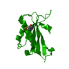
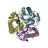
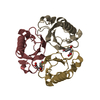
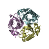
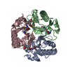
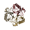
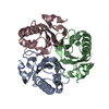
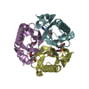
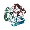
| Title | CRYSTAL STRUCTURE ANALYSIS OF CHORISMATE MUTASE MUTANT C88S/R90K | ||||||
 Components Components | PROTEIN (CHORISMATE MUTASE) | ||||||
 Keywords Keywords | ISOMERASE / chorismate mutase / protein / mutant / pseudo-alpha beta-barrel / trimer | ||||||
| Function / homology |  Function and homology information Function and homology informationchorismate metabolic process / chorismate mutase / chorismate mutase activity / aromatic amino acid biosynthetic process / amino acid biosynthetic process / cytoplasm Similarity search - Function | ||||||
| Biological species |  | ||||||
| Method |  X-RAY DIFFRACTION / MOLECULAR REPLACEMEN / Resolution: 1.9 Å X-RAY DIFFRACTION / MOLECULAR REPLACEMEN / Resolution: 1.9 Å | ||||||
 Authors Authors | Kast, P. / Grisostomi, C. / Chen, I.A. / Li, S. / Krengel, U. / Xue, Y. / Hilvert, D. | ||||||
 Citation Citation | Journal: J.Biol.Chem. / Year: 2000 Title: A strategically positioned cation is crucial for efficient catalysis by chorismate mutase. Authors: Kast, P. / Grisostomi, C. / Chen, I.A. / Li, S. / Krengel, U. / Xue, Y. / Hilvert, D. #1: Journal: Proc.Natl.Acad.Sci.USA / Year: 1996Title: Exploring the active site of chorismate mutase by combinatorial mutagenesis and selection: the importance of electrostatic catalysis Authors: Kast, P. / Asif-Ullah, M. / Jiang, N. / Hilvert, D. #2: Journal: J.Am.Chem.Soc. / Year: 1999Title: Heavy atom isotope effects reveal a highly polarized transition state for chorismate mutase Authors: Gustin, D.J. / Mattei, P. / Kast, P. / Wiest, O. / Lee, L. / Cleland, W.W. / Hilvert, D. | ||||||
| History |
|
- Structure visualization
| Structure viewer | Molecule: MolmilJmol/JSmol |
|---|
- Downloads & links
-Download
| PDBx/mmCIF format | 1fnj.cif.gz | 36.9 KB | Display | PDBx/mmCIF format |
|---|---|---|---|---|
| PDB format | pdb1fnj.ent.gz | 24.6 KB | Display | PDB format |
| PDBx/mmJSON format | 1fnj.json.gz | Tree view | PDBx/mmJSON format | |
| Others |  Other downloads Other downloads |
-Validation report
| Arichive directory | https://data.pdbj.org/pub/pdb/validation_reports/fn/1fnjftp://data.pdbj.org/pub/pdb/validation_reports/fn/1fnj | HTTPS FTP |
|---|
-Related structure data
| Related structure data |  1fnkC 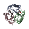 2chtS S: Starting model for refinement C: citing same article ( |
|---|---|
| Similar structure data |

-Links
 PDBj
PDBj
- Assembly
| Deposited unit | 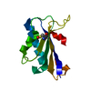
| ||||||||
|---|---|---|---|---|---|---|---|---|---|
| 1 | 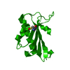
| ||||||||
| Unit cell |
|
-Components
| #1: Protein | Mass: 14479.837 Da / Num. of mol.: 1 / Mutation: YES Source method: isolated from a genetically manipulated source Source: (gene. exp.) Plasmid details: PET-22B(+) FROM NOVAGEN (MADISON, WI) AND PKET3-W, A PET-22B(+) DERIVATIVE ALLOWING FOR T7 PROMOTOR-DRIVEN GENE EXPRESSION Plasmid: PET-22B(+),PKET3-W / Production host: |
|---|---|
| #2: Water | ChemComp-HOH /  Mass: 18.015 Da / Num. of mol.: 27 / Source method: isolated from a natural source / Formula: H2O Mass: 18.015 Da / Num. of mol.: 27 / Source method: isolated from a natural source / Formula: H2O |
| Has protein modification | Y |
-Experimental details
-Experiment
| Experiment | Method: X-RAY DIFFRACTION / Number of used crystals: 1 |
|---|
- Sample preparation
| Crystal | Density Matthews: 2.2 Å3/Da / Density % sol: 45 % | |||||||||||||||||||||||||||||||||||
|---|---|---|---|---|---|---|---|---|---|---|---|---|---|---|---|---|---|---|---|---|---|---|---|---|---|---|---|---|---|---|---|---|---|---|---|---|
| Crystal grow | Temperature: 295 K / Method: vapor diffusion, hanging drop / pH: 7 Details: Protein solution: 10 mM Tris-HCl, 2mM DTT, 0.125 mM EDTA, Reservoir solution: 30% PEG 400, 50 mM Tris-HCl, 50 mM Magnesium Chloride, pH 7.0, VAPOR DIFFUSION, HANGING DROP, temperature 295K | |||||||||||||||||||||||||||||||||||
| Crystal grow | *PLUS PH range low: 7.5 / PH range high: 7 | |||||||||||||||||||||||||||||||||||
| Components of the solutions | *PLUS
|
-Data collection
| Diffraction | Mean temperature: 295 K |
|---|---|
| Diffraction source | Source: ROTATING ANODE / Type: RIGAKU RU200 / Wavelength: 1.54 |
| Detector | Type: RIGAKU RAXIS IIC / Detector: IMAGE PLATE / Date: Nov 1, 1996 |
| Radiation | Protocol: SINGLE WAVELENGTH / Monochromatic (M) / Laue (L): M / Scattering type: x-ray |
| Radiation wavelength | Wavelength: 1.54 Å / Relative weight: 1 |
| Reflection | Resolution: 1.75→30 Å / Num. obs: 10748 / % possible obs: 97.7 % / Observed criterion σ(I): 0 / Redundancy: 4.9 % / Rsym value: 0.06 / Net I/σ(I): 29 |
| Reflection shell | Resolution: 1.75→1.81 Å / Redundancy: 2.5 % / Rsym value: 0.068 / % possible all: 82.1 |
| Reflection | *PLUS Highest resolution: 1.75 Å / Lowest resolution: 30 Å / Rmerge(I) obs: 0.06 |
- Processing
| Software |
| ||||||||||||||||||||||||||||||||||||||||||||||||||||||||||||
|---|---|---|---|---|---|---|---|---|---|---|---|---|---|---|---|---|---|---|---|---|---|---|---|---|---|---|---|---|---|---|---|---|---|---|---|---|---|---|---|---|---|---|---|---|---|---|---|---|---|---|---|---|---|---|---|---|---|---|---|---|---|
| Refinement | Method to determine structure: MOLECULAR REPLACEMEN Starting model: PDB# 2CHT Resolution: 1.9→30 Å / Cross valid method: R-FREE / σ(F): 2
| ||||||||||||||||||||||||||||||||||||||||||||||||||||||||||||
| Refinement step | Cycle: LAST / Resolution: 1.9→30 Å
| ||||||||||||||||||||||||||||||||||||||||||||||||||||||||||||
| Refine LS restraints |
| ||||||||||||||||||||||||||||||||||||||||||||||||||||||||||||
| LS refinement shell | Resolution: 1.9→1.99 Å / Total num. of bins used: 8
| ||||||||||||||||||||||||||||||||||||||||||||||||||||||||||||
| Xplor file | Serial no: 1 / Param file: PARHCSDX.PRO / Topol file: TOPHCSDX.PRO | ||||||||||||||||||||||||||||||||||||||||||||||||||||||||||||
| Refinement | *PLUS Highest resolution: 1.9 Å / Lowest resolution: 30 Å / % reflection Rfree: 5 % | ||||||||||||||||||||||||||||||||||||||||||||||||||||||||||||
| Solvent computation | *PLUS | ||||||||||||||||||||||||||||||||||||||||||||||||||||||||||||
| Displacement parameters | *PLUS | ||||||||||||||||||||||||||||||||||||||||||||||||||||||||||||
| Refine LS restraints | *PLUS
| ||||||||||||||||||||||||||||||||||||||||||||||||||||||||||||
| LS refinement shell | *PLUS Rfactor Rfree: 0.343 / % reflection Rfree: 3.4 % / Rfactor Rwork: 0.316 |
