 Movie
Movie Controller

[English] 日本語
 Yorodumi
- PDB-1fmf: REFINED SOLUTION STRUCTURE OF THE (13C,15N-LABELED) B12-BINDING S... -
+ Open data

- Basic information
| Entry | Database: PDB / ID: 1fmf | ||||||
|---|---|---|---|---|---|---|---|
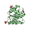
| Title | REFINED SOLUTION STRUCTURE OF THE (13C,15N-LABELED) B12-BINDING SUBUNIT OF GLUTAMATE MUTASE FROM CLOSTRIDIUM TETANOMORPHUM | ||||||
 Components Components | METHYLASPARTATE MUTASE S CHAIN | ||||||
 Keywords Keywords | ISOMERASE / nucleotide binding fold / Rossmann fold | ||||||
| Function / homology |  Function and homology information Function and homology informationmethylaspartate mutase / : / methylaspartate mutase activity / : / cobalamin binding / metal ion binding Similarity search - Function | ||||||
| Biological species |  Clostridium tetanomorphum (bacteria) Clostridium tetanomorphum (bacteria) | ||||||
| Method | SOLUTION NMR / distance geometry, simulated annealing, simulated annealing refinement, energy minimization | ||||||
 Authors Authors | Hoffmann, B. / Konrat, R. / Tollinger, M. / Huhta, M. / Marsh, E.N.G. / Kraeutler, B. | ||||||
 Citation Citation | Journal: Chembiochem / Year: 2001 Title: A protein pre-organized to trap the nucleotide moiety of coenzyme B(12): refined solution structure of the B(12)-binding subunit of glutamate mutase from Clostridium tetanomorphum. Authors: Hoffmann, B. / Tollinger, M. / Konrat, R. / Huhta, M. / Marsh, E.N. / Krautler, B. #1: Journal: Structure / Year: 1998Title: How a protein prepares for B12-binding: structure and dynamics of the B12-binding subunit of glutamate mutase from Clostridium tetanomorphum Authors: Tollinger, M. / Konrat, R. / Hilbert, B.H. / Marsh, E.N.G. / Kraeutler, B. #2: Journal: J.Biol.Chem. / Year: 1994Title: Adenosylcobalamin-dependent glutamate mutase from Clostridium tetanomorphum. Overexpression in Escherichia coli, purification and Characterization of the recombinant enzyme Authors: Holloway, D.E. / Marsh, E.N.G. | ||||||
| History |
|
- Structure visualization
| Structure viewer | Molecule: MolmilJmol/JSmol |
|---|
- Downloads & links
-Download
| PDBx/mmCIF format | 1fmf.cif.gz | 1.2 MB | Display | PDBx/mmCIF format |
|---|---|---|---|---|
| PDB format | pdb1fmf.ent.gz | 997.3 KB | Display | PDB format |
| PDBx/mmJSON format | 1fmf.json.gz | Tree view | PDBx/mmJSON format | |
| Others |  Other downloads Other downloads |
-Validation report
| Arichive directory | https://data.pdbj.org/pub/pdb/validation_reports/fm/1fmfftp://data.pdbj.org/pub/pdb/validation_reports/fm/1fmf | HTTPS FTP |
|---|
-Related structure data
| Related structure data | |
|---|---|
| Similar structure data |


-Links
 PDBj
PDBj- Assembly
| Deposited unit | 
| |||||||||
|---|---|---|---|---|---|---|---|---|---|---|
| 1 |
| |||||||||
| NMR ensembles |
|
-Components
| #1: Protein | Mass: 14763.856 Da / Num. of mol.: 1 Source method: isolated from a genetically manipulated source Source: (gene. exp.) Clostridium tetanomorphum (bacteria) / Plasmid: PMUTSX / Production host: |
|---|
-Experimental details
-Experiment
| Experiment | Method: SOLUTION NMR | ||||||||||||||||
|---|---|---|---|---|---|---|---|---|---|---|---|---|---|---|---|---|---|
| NMR experiment |
| ||||||||||||||||
| NMR details | Text: The structure was determined using triple-resonance NMR spectroscopy. |
- Sample preparation
| Details | Contents: 1.5mM MutS U-98% 15N,13C; 11mM phosphate buffer; / Solvent system: 90% H2O/10% D2O |
|---|---|
| Sample conditions | Ionic strength: 11mM KXH3-XPO4 / pH: 6 / Pressure: 1 atm / Temperature: 299 K |
| Crystal grow | *PLUS Method: other / Details: NMR |
-NMR measurement
| NMR spectrometer | Type: Varian UNITYPLUS / Manufacturer: Varian / Model: UNITYPLUS / Field strength: 500 MHz |
|---|
- Processing
| NMR software |
| ||||||||||||||||||||||||
|---|---|---|---|---|---|---|---|---|---|---|---|---|---|---|---|---|---|---|---|---|---|---|---|---|---|
| Refinement | Method: distance geometry, simulated annealing, simulated annealing refinement, energy minimization Software ordinal: 1 Details: The MutS structure models 1-15 are based on a total of 1792 restraints, 1553 are NOE-derived distance constraints, 184 dihedral angle restraints, 55 distance restraints from hydrogen bonds. ...Details: The MutS structure models 1-15 are based on a total of 1792 restraints, 1553 are NOE-derived distance constraints, 184 dihedral angle restraints, 55 distance restraints from hydrogen bonds. Backbone dihedral angles Phi and Psi were obtained by employing TALOS software [G. Cornilescu et al., J. Biomol. NMR 1999, 13, 289-302]. Phi and Psi torsion angle restraints for the MutS residues 23-30, which form a "nascent" helix [M. Tollinger et al., Structure 1998, 6, 1021-1033], were not used in structure calculations for MutS models 16-30. | ||||||||||||||||||||||||
| NMR representative | Selection criteria: fewest violations | ||||||||||||||||||||||||
| NMR ensemble | Conformer selection criteria: structures with acceptable covalent geometry,structures with favorable non-bond energy,structures with the least restraint violations,structures with the lowest energy Conformers calculated total number: 200 / Conformers submitted total number: 30 |
 NMRPipe