[English] 日本語
Yorodumi
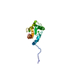
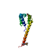
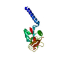
- PDB-1fi5: NMR STRUCTURE OF THE C TERMINAL DOMAIN OF CARDIAC TROPONIN C BOUN... -

+
Open data


ID or keywords:

Loading...

-
Basic information

Entry
Database: PDB / ID: 1fi5
TitleNMR STRUCTURE OF THE C TERMINAL DOMAIN OF CARDIAC TROPONIN C BOUND TO THE N TERMINAL DOMAIN OF CARDIAC TROPONIN I.
ComponentsPROTEIN (TROPONIN C)
KeywordsCONTRACTILE PROTEIN / TROPONIN C-TROPONIN I INTERACTION / CARDIAC / MUSCLE PROTEIN / CALCIUM BINDING PROTEIN
Function / homology
Function and homology information


cardiac Troponin complex / Striated Muscle Contraction / myosin II complex / troponin I binding / skeletal muscle contraction / cardiac muscle contraction / calcium-dependent protein binding / calcium ion binding
Similarity search - Function
EF hand / EF-hand domain pair / EF-hand / Recoverin; domain 1 / EF-hand domain pair / EF-hand, calcium binding motif / EF-Hand 1, calcium-binding site / EF-hand calcium-binding domain. / EF-hand calcium-binding domain profile. / EF-hand domain ...EF hand / EF-hand domain pair / EF-hand / Recoverin; domain 1 / EF-hand domain pair / EF-hand, calcium binding motif / EF-Hand 1, calcium-binding site / EF-hand calcium-binding domain. / EF-hand calcium-binding domain profile. / EF-hand domain / EF-hand domain pair / Orthogonal Bundle / Mainly Alpha
Similarity search - Domain/homology
Troponin C, slow skeletal and cardiac muscles
Similarity search - Component
Biological speciesGallus gallus (chicken)
MethodSOLUTION NMR / DGSA
AuthorsGasmi-Seabrook, G.M. / Howarth, J.W. / Finley, N. / Abusamhadneh, E. / Gaponenko, V. / Brito, R.M. / Solaro, R.J. / Rosevear, P.R.
CitationJournal: Biochemistry / Year: 1999
Title: Solution structures of the C-terminal domain of cardiac troponin C free and bound to the N-terminal domain of cardiac troponin I.
Authors: Gasmi-Seabrook, G.M. / Howarth, J.W. / Finley, N. / Abusamhadneh, E. / Gaponenko, V. / Brito, R.M. / Solaro, R.J. / Rosevear, P.R.
History
DepositionAug 3, 2000Deposition site: RCSB / Processing site: RCSB
SupersessionAug 23, 2000ID: 1GGS
Revision 1.0Aug 23, 2000Provider: repository / Type: Initial release
Revision 1.1Apr 27, 2008Group: Version format compliance
Revision 1.2Jul 13, 2011Group: Version format compliance
Revision 1.3Feb 23, 2022Group: Database references / Derived calculations
Category: database_2 / pdbx_struct_assembly ...database_2 / pdbx_struct_assembly / pdbx_struct_conn_angle / pdbx_struct_oper_list / struct_conn / struct_site
Item: _database_2.pdbx_DOI / _database_2.pdbx_database_accession ..._database_2.pdbx_DOI / _database_2.pdbx_database_accession / _pdbx_struct_conn_angle.ptnr1_auth_comp_id / _pdbx_struct_conn_angle.ptnr1_auth_seq_id / _pdbx_struct_conn_angle.ptnr1_label_atom_id / _pdbx_struct_conn_angle.ptnr1_label_comp_id / _pdbx_struct_conn_angle.ptnr1_label_seq_id / _pdbx_struct_conn_angle.ptnr3_auth_comp_id / _pdbx_struct_conn_angle.ptnr3_auth_seq_id / _pdbx_struct_conn_angle.ptnr3_label_atom_id / _pdbx_struct_conn_angle.ptnr3_label_comp_id / _pdbx_struct_conn_angle.ptnr3_label_seq_id / _pdbx_struct_conn_angle.value / _struct_conn.pdbx_dist_value / _struct_conn.ptnr1_auth_comp_id / _struct_conn.ptnr1_auth_seq_id / _struct_conn.ptnr1_label_asym_id / _struct_conn.ptnr1_label_atom_id / _struct_conn.ptnr1_label_comp_id / _struct_conn.ptnr1_label_seq_id / _struct_conn.ptnr2_auth_comp_id / _struct_conn.ptnr2_auth_seq_id / _struct_conn.ptnr2_label_asym_id / _struct_conn.ptnr2_label_atom_id / _struct_conn.ptnr2_label_comp_id / _struct_conn.ptnr2_label_seq_id / _struct_site.pdbx_auth_asym_id / _struct_site.pdbx_auth_comp_id / _struct_site.pdbx_auth_seq_id
Revision 1.4May 22, 2024Group: Data collection / Category: chem_comp_atom / chem_comp_bond

-
Structure visualization

Structure viewerMolecule:
MolmilJmol/JSmol

Downloads & links

-
Assembly

Deposited unit
A: PROTEIN (TROPONIN C)
hetero molecules


Theoretical massNumber of molelcules
Total (without water)9,5443
Polymers9,4641
Non-polymers802
Water00
1


  • Idetical with deposited unit
  • defined by author
TypeNameSymmetry operationNumber
identity operation1_555x,y,z1
NMR ensembles
DataCriteria
Number of conformers (submitted / calculated)20 / 500SMALLEST RMSD TO AVERAGE STRUCTURE
RepresentativeModel #5closest to the average

-
Components

#1: Protein PROTEIN (TROPONIN C)


Mass: 9463.531 Da / Num. of mol.: 1 / Fragment: RESIDUES 81 - 161
Source method: isolated from a genetically manipulated source
Source: (gene. exp.) Gallus gallus (chicken) / Tissue: MUSCLE
Description: C TERMINAL DOMAIN OF CARDIAC TROPONIN C (81- 161) BOUND TO THE N TERMINAL DOMAIN OF CARDIAC TROPONIN I (33-80), CALCIUM IONS BOUND AT SITES III AND IV
Cell: MYOCYTE / Cellular location: THIN FILAMENT / Organ: HEART / Plasmid: PET-23D+ / Species (production host): Escherichia coli / Gene (production host): CTNC(81-161) / Production host: Escherichia coli BL21(DE3) (bacteria) / Strain (production host): BL21(DE3) / References: UniProt: P09860
#2: Chemical ChemComp-CA / CALCIUM ION


Mass: 40.078 Da / Num. of mol.: 2 / Source method: obtained synthetically / Formula: Ca

-
Experimental details

-
Experiment

ExperimentMethod: SOLUTION NMR
NMR experiment
Conditions-IDExperiment-IDSolution-IDType
11115N-HSQC
12115N-NOESY- HSQC
13115N-TOCSY-HSQC
141HNCO
151HN(CA)CB
161(HB)CBCA (CO)NNH
171H(CCO)NH
181C (CO)NH
191(HB)CB(CGCD)HD
1101(HB)CB(CGCDCE)HE
1111CN- NOESY-HSQC
1121HNHA
1131HNHB.
NMR detailsText: BEST 20 STRUCTURES. THESE STRUCTURES WERE DETERMINED USING TRIPLE-RESONANCE NMR SPECTROSCOPY ON CA2+_SATURATED 15N, 13C-CTNC(81-161) BOUND TO CTNI(33-80).

-
Sample preparation

DetailsContents: 1mM [15N,13C]cTnC(81-161)/cTnI(33-80); 20mM Tris-d11; 100mM KCl; 20mM DTT; 15mM CaCl2; 0.1mM Leupetin; 0.1mM pefabloc; 90% H2O/10% D2O
Solvent system: 90% H2O/10% D2O
Sample conditionsIonic strength: 100mM KCL,15mM CACL2, 100mM KCL,15mM CACL2 / pH: 6.80 / Pressure: ambient / Temperature: 318.00 K
Crystal grow
*PLUS
Method: other / Details: NMR

-
NMR measurement

NMR spectrometer
TypeManufacturerModelField strength (MHz)Spectrometer-ID
Varian INOVA600VarianINOVA6006001
Varian INOVA800VarianINOVA8008002

-
Processing

NMR software
NameVersionDeveloperClassification
CNS1BRUNGERrefinement
CNS1BRUNGERstructure solution
RefinementMethod: DGSA / Software ordinal: 1
Details: REFINEMENT DETAILS CAN BE FOUND IN THE JRNL CITATION ABOVE
NMR representativeSelection criteria: closest to the average
NMR ensembleConformer selection criteria: SMALLEST RMSD TO AVERAGE STRUCTURE
Conformers calculated total number: 500 / Conformers submitted total number: 20

+
About Yorodumi

-
News

-
Feb 9, 2022. New format data for meta-information of EMDB entries

New format data for meta-information of EMDB entries

  • Version 3 of the EMDB header file is now the official format.
  • The previous official version 1.9 will be removed from the archive.

Related info.:EMDB header

External links:wwPDB to switch to version 3 of the EMDB data model

-
Aug 12, 2020. Covid-19 info

Covid-19 info

URL: https://pdbjlvh1.pdbj.org/emnavi/covid19.php

New page: Covid-19 featured information page in EM Navigator.

Related info.:Covid-19 info / Mar 5, 2020. Novel coronavirus structure data

+
Mar 5, 2020. Novel coronavirus structure data

Novel coronavirus structure data

Related info.:Yorodumi Speices / Aug 12, 2020. Covid-19 info

External links:COVID-19 featured content - PDBj / Molecule of the Month (242):Coronavirus Proteases

+
Jan 31, 2019. EMDB accession codes are about to change! (news from PDBe EMDB page)

EMDB accession codes are about to change! (news from PDBe EMDB page)

  • The allocation of 4 digits for EMDB accession codes will soon come to an end. Whilst these codes will remain in use, new EMDB accession codes will include an additional digit and will expand incrementally as the available range of codes is exhausted. The current 4-digit format prefixed with “EMD-” (i.e. EMD-XXXX) will advance to a 5-digit format (i.e. EMD-XXXXX), and so on. It is currently estimated that the 4-digit codes will be depleted around Spring 2019, at which point the 5-digit format will come into force.
  • The EM Navigator/Yorodumi systems omit the EMD- prefix.

Related info.:Q: What is EMD? / ID/Accession-code notation in Yorodumi/EM Navigator

External links:EMDB Accession Codes are Changing Soon! / Contact to PDBj

+
Jul 12, 2017. Major update of PDB

Major update of PDB

  • wwPDB released updated PDB data conforming to the new PDBx/mmCIF dictionary.
  • This is a major update changing the version number from 4 to 5, and with Remediation, in which all the entries are updated.
  • In this update, many items about electron microscopy experimental information are reorganized (e.g. em_software).
  • Now, EM Navigator and Yorodumi are based on the updated data.

External links:wwPDB Remediation / Enriched Model Files Conforming to OneDep Data Standards Now Available in the PDB FTP Archive

-
Yorodumi

Thousand views of thousand structures

  • Yorodumi is a browser for structure data from EMDB, PDB, SASBDB, etc.
  • This page is also the successor to EM Navigator detail page, and also detail information page/front-end page for Omokage search.
  • The word "yorodu" (or yorozu) is an old Japanese word meaning "ten thousand". "mi" (miru) is to see.

Related info.:EMDB / PDB / SASBDB / Comparison of 3 databanks / Yorodumi Search / Aug 31, 2016. New EM Navigator & Yorodumi / Yorodumi Papers / Jmol/JSmol / Function and homology information / Changes in new EM Navigator and Yorodumi

Read more