 Movie
Movie Controller

[English] 日本語
 Yorodumi
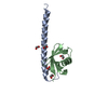
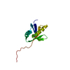
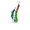
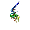
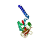
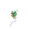
- PDB-1scv: NMR STRUCTURE OF THE C TERMINAL DOMAIN OF CARDIAC TROPONIN C BOUN... -
+ Open data

- Basic information
| Entry | Database: PDB / ID: 1scv | ||||||
|---|---|---|---|---|---|---|---|
| Title | NMR STRUCTURE OF THE C TERMINAL DOMAIN OF CARDIAC TROPONIN C BOUND TO THE N TERMINAL DOMAIN OF CARDIAC TROPONIN I | ||||||
 Components Components | Troponin C, slow skeletal and cardiac muscles | ||||||
 Keywords Keywords | CONTRACTILE PROTEIN / STRUCTURAL PROTEIN / TROPONIN C-TROPONIN I INTERACTION / CARDIAC / MUSCLE PROTEIN / CALCIUM BINDING PROTEIN | ||||||
| Function / homology |  Function and homology information Function and homology informationcardiac Troponin complex / Striated Muscle Contraction / troponin I binding / skeletal muscle contraction / cardiac muscle contraction / calcium-dependent protein binding / calcium ion binding Similarity search - Function | ||||||
| Biological species |  | ||||||
| Method | SOLUTION NMR / DGSA | ||||||
 Authors Authors | Finley, N.L. / Howarth, J.W. / Rosevear, P.R. | ||||||
 Citation Citation | Journal: Biochemistry / Year: 2004 Title: Structure of the Mg2+-loaded C-lobe of cardiac troponin C bound to the N-domain of cardiac troponin I: comparison with the Ca2+-loaded structure. Authors: Finley, N.L. / Howarth, J.W. / Rosevear, P.R. #1: Journal: Biochemistry / Year: 1999Title: Solution structures of the C-terminal domain of cardiac troponin C free and bound to the N-terminal domain of cardiac troponin I Authors: Gasmi-Seabrook, G.M. / Howarth, J.W. / Finley, N. / Abusamhadneh, E. / Gaponenko, V. / Brito, R.M. / Solaro, R.J. / Rosevear, P.R. | ||||||
| History |
| ||||||
| Remark 650 | HELIX DETERMINATION METHOD: AUTHOR | ||||||
| Remark 700 | SHEET DETERMINATION METHOD: AUTHOR |
- Structure visualization
| Structure viewer | Molecule: MolmilJmol/JSmol |
|---|
- Downloads & links
-Download
| PDBx/mmCIF format | 1scv.cif.gz | 502.1 KB | Display | PDBx/mmCIF format |
|---|---|---|---|---|
| PDB format | pdb1scv.ent.gz | 419.9 KB | Display | PDB format |
| PDBx/mmJSON format | 1scv.json.gz | Tree view | PDBx/mmJSON format | |
| Others |  Other downloads Other downloads |
-Validation report
| Arichive directory | https://data.pdbj.org/pub/pdb/validation_reports/sc/1scvftp://data.pdbj.org/pub/pdb/validation_reports/sc/1scv | HTTPS FTP |
|---|
-Related structure data


-Links
 PDBj
PDBj

- Assembly
| Deposited unit | 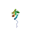
| |||||||||
|---|---|---|---|---|---|---|---|---|---|---|
| 1 |
| |||||||||
| NMR ensembles |
|
-Components
| #1: Protein | Mass: 9463.531 Da / Num. of mol.: 1 / Fragment: C-terminal domain (RESIDUES 81 - 161) Source method: isolated from a genetically manipulated source Source: (gene. exp.)  |
|---|---|
| #2: Chemical |   Mass: 40.078 Da / Num. of mol.: 2 / Source method: obtained synthetically / Formula: Ca Mass: 40.078 Da / Num. of mol.: 2 / Source method: obtained synthetically / Formula: Ca |
-Experimental details
-Experiment
| Experiment | Method: SOLUTION NMR | ||||||||||||||||||||||||||||
|---|---|---|---|---|---|---|---|---|---|---|---|---|---|---|---|---|---|---|---|---|---|---|---|---|---|---|---|---|---|
| NMR experiment |
| ||||||||||||||||||||||||||||
| NMR details | Text: BEST 20 STRUCTURES. THESE STRUCTURES WERE DETERMINED USING TRIPLE-RESONANCE NMR SPECTROSCOPY ON CA2+_SATURATED 15N, 13C-CTNC(81-161) BOUND TO CTNI(33-80). |
 HSQC
HSQC- Sample preparation
| Details | Contents: 1MM [15N,13C]CTNC(81-161)/CTNI(33-80), 20MM TRIS-D11, 100MM KCL, 20MM DTT, 15MM CACL2, 0.1MM LEUPETIN, 0.1MM PEFABLOC; 90% H2O, 10% D2O Solvent system: 90% H2O/10% D2O |
|---|---|
| Sample conditions | Ionic strength: 100mM KCL, 15mM CACL2 / pH: 6.8 / Pressure: AMBIENT / Temperature: 318.00 K |
-NMR measurement
| Radiation | Protocol: SINGLE WAVELENGTH / Monochromatic (M) / Laue (L): M | |||||||||||||||
|---|---|---|---|---|---|---|---|---|---|---|---|---|---|---|---|---|
| Radiation wavelength | Relative weight: 1 | |||||||||||||||
| NMR spectrometer |
|
- Processing
| NMR software |
| ||||||||||||||||
|---|---|---|---|---|---|---|---|---|---|---|---|---|---|---|---|---|---|
| Refinement | Method: DGSA / Software ordinal: 1 Details: The structures are based on a total of 926 restraints, 787 are NOE-derived distance restraints, 91 dihedral angle restraints, 4 distance restraints from hydrogen bonds and 44 dipolar coupling restraints. | ||||||||||||||||
| NMR representative | Selection criteria: lowest energy | ||||||||||||||||
| NMR ensemble | Conformer selection criteria: structures with the lowest energy Conformers calculated total number: 20 / Conformers submitted total number: 20 |
