 Movie
Movie Controller

[English] 日本語
 Yorodumi
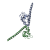
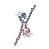
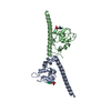
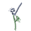
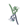
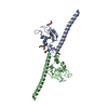
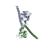
- PDB-1f3h: X-RAY CRYSTAL STRUCTURE OF THE HUMAN ANTI-APOPTOTIC PROTEIN SURVIVIN -
+ Open data

- Basic information
| Entry | Database: PDB / ID: 1f3h | ||||||
|---|---|---|---|---|---|---|---|
| Title | X-RAY CRYSTAL STRUCTURE OF THE HUMAN ANTI-APOPTOTIC PROTEIN SURVIVIN | ||||||
 Components Components | SURVIVIN | ||||||
 Keywords Keywords | APOPTOSIS / APOPTOSIS INHIBITOR SURVIVIN / THIOL PROTEASE INHIBITOR / ALPHA-BETA | ||||||
| Function / homology |  Function and homology information Function and homology informationsurvivin complex / : / positive regulation of mitotic sister chromatid separation / positive regulation of mitotic cytokinesis / positive regulation of mitotic cell cycle spindle assembly checkpoint / positive regulation of exit from mitosis / mitotic spindle midzone assembly / positive regulation of attachment of mitotic spindle microtubules to kinetochore / interphase microtubule organizing center / chromosome passenger complex ...survivin complex / : / positive regulation of mitotic sister chromatid separation / positive regulation of mitotic cytokinesis / positive regulation of mitotic cell cycle spindle assembly checkpoint / positive regulation of exit from mitosis / mitotic spindle midzone assembly / positive regulation of attachment of mitotic spindle microtubules to kinetochore / interphase microtubule organizing center / chromosome passenger complex / protein-containing complex localization / cysteine-type endopeptidase inhibitor activity involved in apoptotic process / cobalt ion binding / nuclear chromosome / mitotic spindle assembly checkpoint signaling / spindle midzone / TP53 regulates transcription of several additional cell death genes whose specific roles in p53-dependent apoptosis remain uncertain / mitotic cytokinesis / SUMOylation of DNA replication proteins / chromosome, centromeric region / mitotic spindle assembly / cysteine-type endopeptidase inhibitor activity / cytoplasmic microtubule / Amplification of signal from unattached kinetochores via a MAD2 inhibitory signal / Mitotic Prometaphase / EML4 and NUDC in mitotic spindle formation / centriole / tubulin binding / positive regulation of mitotic cell cycle / Resolution of Sister Chromatid Cohesion / mitotic spindle organization / spindle microtubule / chromosome segregation / RHO GTPases Activate Formins / sensory perception of sound / kinetochore / small GTPase binding / G2/M transition of mitotic cell cycle / Separation of Sister Chromatids / mitotic cell cycle / protein-folding chaperone binding / Neddylation / microtubule cytoskeleton / midbody / Interleukin-4 and Interleukin-13 signaling / microtubule binding / microtubule / protein heterodimerization activity / cell division / negative regulation of DNA-templated transcription / positive regulation of cell population proliferation / apoptotic process / negative regulation of apoptotic process / enzyme binding / protein homodimerization activity / protein-containing complex / zinc ion binding / nucleoplasm / metal ion binding / identical protein binding / nucleus / cytoplasm / cytosol Similarity search - Function | ||||||
| Biological species |  Homo sapiens (human) Homo sapiens (human) | ||||||
| Method |  X-RAY DIFFRACTION / SYNCHROTRON / Resolution: 2.58 Å X-RAY DIFFRACTION / SYNCHROTRON / Resolution: 2.58 Å | ||||||
 Authors Authors | Verdecia, M.A. / Huang, H. / Dutil, E. / Hunter, T. / Noel, J.P. | ||||||
 Citation Citation | Journal: Nat.Struct.Biol. / Year: 2000 Title: Structure of the human anti-apoptotic protein survivin reveals a dimeric arrangement. Authors: Verdecia, M.A. / Huang, H. / Dutil, E. / Kaiser, D.A. / Hunter, T. / Noel, J.P. | ||||||
| History |
|
- Structure visualization
| Structure viewer | Molecule: MolmilJmol/JSmol |
|---|
- Downloads & links
-Download
| PDBx/mmCIF format | 1f3h.cif.gz | 70.8 KB | Display | PDBx/mmCIF format |
|---|---|---|---|---|
| PDB format | pdb1f3h.ent.gz | 53.1 KB | Display | PDB format |
| PDBx/mmJSON format | 1f3h.json.gz | Tree view | PDBx/mmJSON format | |
| Others |  Other downloads Other downloads |
-Validation report
| Summary document | 1f3h_validation.pdf.gz | 389.3 KB | Display | wwPDB validaton report |
|---|---|---|---|---|
| Full document | 1f3h_full_validation.pdf.gz | 408.7 KB | Display | |
| Data in XML | 1f3h_validation.xml.gz | 10.1 KB | Display | |
| Data in CIF | 1f3h_validation.cif.gz | 14.6 KB | Display | |
| Arichive directory | https://data.pdbj.org/pub/pdb/validation_reports/f3/1f3hftp://data.pdbj.org/pub/pdb/validation_reports/f3/1f3h | HTTPS FTP |
-Related structure data
| Similar structure data |
|---|


-Links
 PDBj
PDBj


- Assembly
| Deposited unit | 
| ||||||||
|---|---|---|---|---|---|---|---|---|---|
| 1 |
| ||||||||
| Unit cell |
|
-Components
| #1: Protein | Mass: 16432.773 Da / Num. of mol.: 2 / Mutation: L54M Source method: isolated from a genetically manipulated source Source: (gene. exp.) Homo sapiens (human) / Plasmid: PHIS8-3 / Production host:  #2: Chemical |   Mass: 65.409 Da / Num. of mol.: 2 / Source method: obtained synthetically / Formula: Zn Mass: 65.409 Da / Num. of mol.: 2 / Source method: obtained synthetically / Formula: Zn#3: Chemical |   Mass: 96.063 Da / Num. of mol.: 3 / Source method: obtained synthetically / Formula: SO4 Mass: 96.063 Da / Num. of mol.: 3 / Source method: obtained synthetically / Formula: SO4#4: Water | ChemComp-HOH / |  Mass: 18.015 Da / Num. of mol.: 90 / Source method: isolated from a natural source / Formula: H2O Mass: 18.015 Da / Num. of mol.: 90 / Source method: isolated from a natural source / Formula: H2OHas protein modification | Y | |
|---|
-Experimental details
-Experiment
| Experiment | Method: X-RAY DIFFRACTION / Number of used crystals: 1 |
|---|
- Sample preparation
| Crystal | Density Matthews: 3.9 Å3/Da / Density % sol: 68.47 % | ||||||||||||||||||||||||||||||||||||||||||
|---|---|---|---|---|---|---|---|---|---|---|---|---|---|---|---|---|---|---|---|---|---|---|---|---|---|---|---|---|---|---|---|---|---|---|---|---|---|---|---|---|---|---|---|
| Crystal grow | Temperature: 298 K / Method: vapor diffusion, hanging drop / pH: 7.5 Details: PEG 8000, LITHIUMSULPHATE, HEPES, pH 7.5, VAPOR DIFFUSION, HANGING DROP, temperature 298.0K | ||||||||||||||||||||||||||||||||||||||||||
| Crystal grow | *PLUS Temperature: 4 ℃ | ||||||||||||||||||||||||||||||||||||||||||
| Components of the solutions | *PLUS
|
-Data collection
| Diffraction |
| ||||||||||||||||||||
|---|---|---|---|---|---|---|---|---|---|---|---|---|---|---|---|---|---|---|---|---|---|
| Diffraction source |
| ||||||||||||||||||||
| Detector |
| ||||||||||||||||||||
| Radiation |
| ||||||||||||||||||||
| Radiation wavelength |
| ||||||||||||||||||||
| Reflection | Resolution: 2.58→54.12 Å / Observed criterion σ(F): 2 / Observed criterion σ(I): 2 / Biso Wilson estimate: 60.5 Å2 | ||||||||||||||||||||
| Reflection shell | Resolution: 2.58→2.64 Å / Redundancy: 3.3 % / Rmerge(I) obs: 0.388 / % possible all: 95.5 | ||||||||||||||||||||
| Reflection | *PLUS Num. obs: 15366 / % possible obs: 95.5 % / Num. measured all: 29469 / Rmerge(I) obs: 0.036 | ||||||||||||||||||||
| Reflection shell | *PLUS % possible obs: 92 % / Rmerge(I) obs: 0.349 |

- Processing
| Software |
| ||||||||||||||||||||||||||||||||||||||||||||||||||||||||||||||||||||||||||||||||
|---|---|---|---|---|---|---|---|---|---|---|---|---|---|---|---|---|---|---|---|---|---|---|---|---|---|---|---|---|---|---|---|---|---|---|---|---|---|---|---|---|---|---|---|---|---|---|---|---|---|---|---|---|---|---|---|---|---|---|---|---|---|---|---|---|---|---|---|---|---|---|---|---|---|---|---|---|---|---|---|---|---|
| Refinement | Resolution: 2.58→54.12 Å / Rfactor Rfree error: 0.01 / Data cutoff high absF: 1711716.61 / Data cutoff low absF: 0 / Isotropic thermal model: RESTRAINED / Cross valid method: THROUGHOUT / σ(F): 0 / σ(I): 2 / Stereochemistry target values: ENGH & HUBER
| ||||||||||||||||||||||||||||||||||||||||||||||||||||||||||||||||||||||||||||||||
| Solvent computation | Solvent model: FLAT MODEL / Bsol: 65.36 Å2 / ksol: 0.3262 e/Å3 | ||||||||||||||||||||||||||||||||||||||||||||||||||||||||||||||||||||||||||||||||
| Displacement parameters | Biso mean: 83.8 Å2
| ||||||||||||||||||||||||||||||||||||||||||||||||||||||||||||||||||||||||||||||||
| Refine analyze |
| ||||||||||||||||||||||||||||||||||||||||||||||||||||||||||||||||||||||||||||||||
| Refinement step | Cycle: LAST / Resolution: 2.58→54.12 Å
| ||||||||||||||||||||||||||||||||||||||||||||||||||||||||||||||||||||||||||||||||
| Refine LS restraints |
| ||||||||||||||||||||||||||||||||||||||||||||||||||||||||||||||||||||||||||||||||
| LS refinement shell | Resolution: 2.58→2.74 Å / Rfactor Rfree error: 0.042 / Total num. of bins used: 6
| ||||||||||||||||||||||||||||||||||||||||||||||||||||||||||||||||||||||||||||||||
| Xplor file |
| ||||||||||||||||||||||||||||||||||||||||||||||||||||||||||||||||||||||||||||||||
| Software | *PLUS Name: CNS / Version: 1 / Classification: refinement | ||||||||||||||||||||||||||||||||||||||||||||||||||||||||||||||||||||||||||||||||
| Refinement | *PLUS σ(F): 0 / % reflection Rfree: 5 % | ||||||||||||||||||||||||||||||||||||||||||||||||||||||||||||||||||||||||||||||||
| Solvent computation | *PLUS | ||||||||||||||||||||||||||||||||||||||||||||||||||||||||||||||||||||||||||||||||
| Displacement parameters | *PLUS Biso mean: 83.8 Å2 | ||||||||||||||||||||||||||||||||||||||||||||||||||||||||||||||||||||||||||||||||
| Refine LS restraints | *PLUS
| ||||||||||||||||||||||||||||||||||||||||||||||||||||||||||||||||||||||||||||||||
| LS refinement shell | *PLUS Rfactor Rfree: 0.448 / % reflection Rfree: 4.7 % / Rfactor Rwork: 0.406 |
