 Movie
Movie Controller

[English] 日本語
 Yorodumi
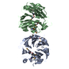
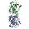
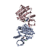
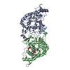
- PDB-1ext: EXTRACELLULAR DOMAIN OF THE 55KDA TUMOR NECROSIS FACTOR RECEPTOR.... -
+ Open data

- Basic information
| Entry | Database: PDB / ID: 1ext | ||||||
|---|---|---|---|---|---|---|---|
| Title | EXTRACELLULAR DOMAIN OF THE 55KDA TUMOR NECROSIS FACTOR RECEPTOR. CRYSTALLIZED AT PH3.7 IN P 21 21 21. | ||||||
 Components Components | TUMOR NECROSIS FACTOR RECEPTOR | ||||||
 Keywords Keywords | SIGNALLING PROTEIN / BINDING PROTEIN / CYTOKINE | ||||||
| Function / homology |  Function and homology information Function and homology information: / pulmonary valve development / tumor necrosis factor receptor superfamily complex / aortic valve development / negative regulation of extracellular matrix constituent secretion / tumor necrosis factor receptor activity / : / positive regulation of apoptotic process involved in morphogenesis / TNFs bind their physiological receptors / tumor necrosis factor binding ...: / pulmonary valve development / tumor necrosis factor receptor superfamily complex / aortic valve development / negative regulation of extracellular matrix constituent secretion / tumor necrosis factor receptor activity / : / positive regulation of apoptotic process involved in morphogenesis / TNFs bind their physiological receptors / tumor necrosis factor binding / negative regulation of cardiac muscle hypertrophy / TNF signaling / regulation of establishment of endothelial barrier / regulation of tumor necrosis factor-mediated signaling pathway / TNFR1-induced proapoptotic signaling / TNFR1-mediated ceramide production / prostaglandin metabolic process / positive regulation of lipid metabolic process / Interleukin-10 signaling / extrinsic apoptotic signaling pathway via death domain receptors / positive regulation of execution phase of apoptosis / cell surface receptor signaling pathway via JAK-STAT / canonical NF-kappaB signal transduction / tumor necrosis factor-mediated signaling pathway / protein localization to plasma membrane / negative regulation of canonical NF-kappaB signal transduction / TNFR1-induced NF-kappa-B signaling pathway / cellular response to mechanical stimulus / Regulation of TNFR1 signaling / negative regulation of inflammatory response / intrinsic apoptotic signaling pathway in response to DNA damage / cytokine-mediated signaling pathway / protein polyubiquitination / positive regulation of inflammatory response / signaling receptor activity / transcription by RNA polymerase II / positive regulation of canonical NF-kappaB signal transduction / signaling receptor complex / defense response to bacterium / membrane raft / inflammatory response / Golgi membrane / cell surface / positive regulation of transcription by RNA polymerase II / : / extracellular region / membrane / plasma membrane Similarity search - Function | ||||||
| Biological species |  Homo sapiens (human) Homo sapiens (human) | ||||||
| Method |  X-RAY DIFFRACTION / SYNCHROTRON / MOLECULAR REPLACEMENT / Resolution: 1.85 Å X-RAY DIFFRACTION / SYNCHROTRON / MOLECULAR REPLACEMENT / Resolution: 1.85 Å | ||||||
 Authors Authors | Naismith, J.H. / Sprang, S.R. | ||||||
 Citation Citation | Journal: Structure / Year: 1996 Title: Structures of the extracellular domain of the type I tumor necrosis factor receptor. Authors: Naismith, J.H. / Devine, T.Q. / Kohno, T. / Sprang, S.R. #1: Journal: J.Biol.Chem. / Year: 1995Title: Crystallographic Evidence for Dimerization of Unliganded Tumor Necrosis Factor Receptor Authors: Naismith, J.H. / Devine, T.Q. / Brandhuber, B.J. / Sprang, S.R. #2: Journal: J.Mol.Biol. / Year: 1994Title: Two Crystal Forms of the Extracellular Domain of Type I Tumor Necrosis Factor Receptor Authors: Rodseth, L.E. / Brandhuber, B. / Devine, T.Q. / Eck, M.J. / Hale, K. / Naismith, J.H. / Sprang, S.R. #3: Journal: Cell(Cambridge,Mass.) / Year: 1993Title: Crystal Structure of the Soluble Human 55 Kd Tnf Receptor-Human Tnf Beta Complex: Implications for Tnf Receptor Activation Authors: Banner, D.W. / D'Arcy, A. / Janes, W. / Gentz, R. / Schoenfeld, H.J. / Broger, C. / Loetscher, H. / Lesslauer, W. | ||||||
| History |
|
- Structure visualization
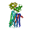
| Structure viewer | Molecule: MolmilJmol/JSmol |
|---|
- Downloads & links
-Download
| PDBx/mmCIF format | 1ext.cif.gz | 85.5 KB | Display | PDBx/mmCIF format |
|---|---|---|---|---|
| PDB format | pdb1ext.ent.gz | 63.1 KB | Display | PDB format |
| PDBx/mmJSON format | 1ext.json.gz | Tree view | PDBx/mmJSON format | |
| Others |  Other downloads Other downloads |
-Validation report
| Arichive directory | https://data.pdbj.org/pub/pdb/validation_reports/ex/1extftp://data.pdbj.org/pub/pdb/validation_reports/ex/1ext | HTTPS FTP |
|---|
-Related structure data
| Related structure data | 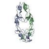 1ncfS S: Starting model for refinement |
|---|---|
| Similar structure data |


-Links
 PDBj
PDBj


- Assembly
| Deposited unit | 
| ||||||||
|---|---|---|---|---|---|---|---|---|---|
| 1 |
| ||||||||
| Unit cell |
|
-Components
| #1: Protein | Mass: 18335.830 Da / Num. of mol.: 2 / Fragment: EXTRACELLULAR DOMAIN Source method: isolated from a genetically manipulated source Source: (gene. exp.) Homo sapiens (human)Description: THE CONSTRUCT CONTAINS RESIDUES 12 TO 172 OF THE MATURE SEQUENCE OF THE ENTIRE RECEPTOR. RESIDUE 11 IS MUTATED TO MET AS A RESULT OF THE EXPRESSION SYSTEM Production host:  #2: Chemical |   Mass: 96.063 Da / Num. of mol.: 3 / Source method: obtained synthetically / Formula: SO4 Mass: 96.063 Da / Num. of mol.: 3 / Source method: obtained synthetically / Formula: SO4#3: Chemical | ChemComp-MG / |   Mass: 24.305 Da / Num. of mol.: 1 / Source method: obtained synthetically / Formula: Mg Mass: 24.305 Da / Num. of mol.: 1 / Source method: obtained synthetically / Formula: Mg#4: Water | ChemComp-HOH / |  Mass: 18.015 Da / Num. of mol.: 400 / Source method: isolated from a natural source / Formula: H2O Mass: 18.015 Da / Num. of mol.: 400 / Source method: isolated from a natural source / Formula: H2OHas protein modification | Y | |
|---|
-Experimental details
-Experiment
| Experiment | Method: X-RAY DIFFRACTION / Number of used crystals: 1 |
|---|
- Sample preparation
| Crystal | Density Matthews: 3.03 Å3/Da / Density % sol: 58 % | |||||||||||||||||||||||||||||||||||||||||||||||||
|---|---|---|---|---|---|---|---|---|---|---|---|---|---|---|---|---|---|---|---|---|---|---|---|---|---|---|---|---|---|---|---|---|---|---|---|---|---|---|---|---|---|---|---|---|---|---|---|---|---|---|
| Crystal grow | pH: 3.7 / Details: CRYSTALLIZED AT PH3.7 IN P 21 21 21. | |||||||||||||||||||||||||||||||||||||||||||||||||
| Crystal | *PLUS | |||||||||||||||||||||||||||||||||||||||||||||||||
| Crystal grow | *PLUS Temperature: 293 K / Method: vapor diffusion, sitting drop / Details: Rodseth, L.E., (1994) J.Mol.Biol., 239, 332. | |||||||||||||||||||||||||||||||||||||||||||||||||
| Components of the solutions | *PLUS
|
-Data collection
| Diffraction | Mean temperature: 123 K |
|---|---|
| Diffraction source | Source: SYNCHROTRON / Site: SRS  / Beamline: PX9.5 / Wavelength: 0.992 / Beamline: PX9.5 / Wavelength: 0.992 |
| Detector | Type: MARRESEARCH / Detector: IMAGE PLATE / Date: Mar 15, 1994 |
| Radiation | Monochromatic (M) / Laue (L): M / Scattering type: x-ray |
| Radiation wavelength | Wavelength: 0.992 Å / Relative weight: 1 |
| Reflection | Num. obs: 36977 / % possible obs: 97 % / Observed criterion σ(I): 0 / Redundancy: 6 % / Rmerge(I) obs: 0.051 |
| Reflection shell | Resolution: 1.85→1.93 Å / % possible all: 77 |
| Reflection | *PLUS Highest resolution: 1.85 Å / Lowest resolution: 11 Å / Num. obs: 37300 |
- Processing
| Software |
| ||||||||||||||||||||||||||||||||||||||||||||||||||||||||||||
|---|---|---|---|---|---|---|---|---|---|---|---|---|---|---|---|---|---|---|---|---|---|---|---|---|---|---|---|---|---|---|---|---|---|---|---|---|---|---|---|---|---|---|---|---|---|---|---|---|---|---|---|---|---|---|---|---|---|---|---|---|---|
| Refinement | Method to determine structure: MOLECULAR REPLACEMENT Starting model: 1NCF Resolution: 1.85→8 Å / Cross valid method: FREE R / σ(F): 0 Details: ATOMS WITH ZERO OCCUPANCY ARE STEREOCHEMICALLY MODELED (68 OUT OF 2162) AND WERE NOT LOCATED IN EXPERIMENTAL MAPS.
| ||||||||||||||||||||||||||||||||||||||||||||||||||||||||||||
| Displacement parameters | Biso mean: 19 Å2 | ||||||||||||||||||||||||||||||||||||||||||||||||||||||||||||
| Refinement step | Cycle: LAST / Resolution: 1.85→8 Å
| ||||||||||||||||||||||||||||||||||||||||||||||||||||||||||||
| Refine LS restraints |
| ||||||||||||||||||||||||||||||||||||||||||||||||||||||||||||
| Software | *PLUS Name: X-PLOR / Version: 3.1 / Classification: refinement | ||||||||||||||||||||||||||||||||||||||||||||||||||||||||||||
| Refinement | *PLUS Rfactor obs: 0.205 | ||||||||||||||||||||||||||||||||||||||||||||||||||||||||||||
| Solvent computation | *PLUS | ||||||||||||||||||||||||||||||||||||||||||||||||||||||||||||
| Displacement parameters | *PLUS | ||||||||||||||||||||||||||||||||||||||||||||||||||||||||||||
| Refine LS restraints | *PLUS
|
