 Movie
Movie Controller

[English] 日本語
 Yorodumi
- PDB-1ex7: CRYSTAL STRUCTURE OF YEAST GUANYLATE KINASE IN COMPLEX WITH GUANO... -
+ Open data

- Basic information
| Entry | Database: PDB / ID: 1ex7 | ||||||
|---|---|---|---|---|---|---|---|
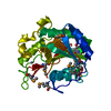
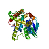
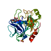
| Title | CRYSTAL STRUCTURE OF YEAST GUANYLATE KINASE IN COMPLEX WITH GUANOSINE-5'-MONOPHOSPHATE | ||||||
 Components Components | GUANYLATE KINASE | ||||||
 Keywords Keywords | TRANSFERASE / GUANYLATE KINASE / SUBSTRATE-INDUCED FIT / DOMAIN MOVEMENT / GMP / ATP / SUBSTRATE SPECIFICITY | ||||||
| Function / homology |  Function and homology information Function and homology informationAzathioprine ADME / GDP biosynthetic process / guanylate kinase / purine nucleotide metabolic process / Interconversion of nucleotide di- and triphosphates / GMP kinase activity / ATP binding / nucleus / cytoplasm / cytosol Similarity search - Function | ||||||
| Biological species |  | ||||||
| Method |  X-RAY DIFFRACTION / REFINEMENT OF THE MODEL / Resolution: 1.9 Å X-RAY DIFFRACTION / REFINEMENT OF THE MODEL / Resolution: 1.9 Å | ||||||
 Authors Authors | Blaszczyk, J. / Ji, X. | ||||||
 Citation Citation | Journal: J.Mol.Biol. / Year: 2001 Title: Crystal structure of unligated guanylate kinase from yeast reveals GMP-induced conformational changes. Authors: Blaszczyk, J. / Li, Y. / Yan, H. / Ji, X. | ||||||
| History |
|
- Structure visualization
| Structure viewer | Molecule: MolmilJmol/JSmol |
|---|
- Downloads & links
-Download
| PDBx/mmCIF format | 1ex7.cif.gz | 56 KB | Display | PDBx/mmCIF format |
|---|---|---|---|---|
| PDB format | pdb1ex7.ent.gz | 39.3 KB | Display | PDB format |
| PDBx/mmJSON format | 1ex7.json.gz | Tree view | PDBx/mmJSON format | |
| Others |  Other downloads Other downloads |
-Validation report
| Summary document | 1ex7_validation.pdf.gz | 773.8 KB | Display | wwPDB validaton report |
|---|---|---|---|---|
| Full document | 1ex7_full_validation.pdf.gz | 777.5 KB | Display | |
| Data in XML | 1ex7_validation.xml.gz | 12.2 KB | Display | |
| Data in CIF | 1ex7_validation.cif.gz | 17.4 KB | Display | |
| Arichive directory | https://data.pdbj.org/pub/pdb/validation_reports/ex/1ex7ftp://data.pdbj.org/pub/pdb/validation_reports/ex/1ex7 | HTTPS FTP |
-Related structure data
| Related structure data |  1ex6C  1gkyS S: Starting model for refinement C: citing same article ( |
|---|---|
| Similar structure data |


-Links
 PDBj
PDBj

- Assembly
| Deposited unit | 
| ||||||||
|---|---|---|---|---|---|---|---|---|---|
| 1 |
| ||||||||
| Unit cell |
|
-Components
| #1: Protein | Mass: 20533.152 Da / Num. of mol.: 1 Source method: isolated from a genetically manipulated source Source: (gene. exp.) Plasmid: PET17B / Species (production host): Escherichia coli / Production host:  | ||||
|---|---|---|---|---|---|
| #2: Chemical |   Mass: 96.063 Da / Num. of mol.: 2 / Source method: obtained synthetically / Formula: SO4 Mass: 96.063 Da / Num. of mol.: 2 / Source method: obtained synthetically / Formula: SO4#3: Chemical | ChemComp-5GP / |   Mass: 363.221 Da / Num. of mol.: 1 / Source method: obtained synthetically / Formula: C10H14N5O8P Mass: 363.221 Da / Num. of mol.: 1 / Source method: obtained synthetically / Formula: C10H14N5O8P#4: Water | ChemComp-HOH / |  Mass: 18.015 Da / Num. of mol.: 208 / Source method: isolated from a natural source / Formula: H2O Mass: 18.015 Da / Num. of mol.: 208 / Source method: isolated from a natural source / Formula: H2O |
-Experimental details
-Experiment
| Experiment | Method: X-RAY DIFFRACTION / Number of used crystals: 1 |
|---|
- Sample preparation
| Crystal | Density Matthews: 2.3 Å3/Da / Density % sol: 44 % | ||||||||||||||||||||||||||||||
|---|---|---|---|---|---|---|---|---|---|---|---|---|---|---|---|---|---|---|---|---|---|---|---|---|---|---|---|---|---|---|---|
| Crystal grow | Temperature: 292 K / Method: vapor diffusion, hanging drop / pH: 5.5 Details: ammonium sulfate, sodium phosphate, pH 5.5, VAPOR DIFFUSION, HANGING DROP, temperature 292K | ||||||||||||||||||||||||||||||
| Crystal grow | *PLUS pH: 7 Details: drop consists of equal amounts of protein and reservoir solutions | ||||||||||||||||||||||||||||||
| Components of the solutions | *PLUS
|
-Data collection
| Diffraction | Mean temperature: 293 K |
|---|---|
| Diffraction source | Source: ROTATING ANODE / Type: ENRAF-NONIUS FR591 / Wavelength: 1.54178 |
| Detector | Type: MAC Science DIP-2000 / Detector: IMAGE PLATE / Date: Dec 5, 1996 / Details: MIRROR |
| Radiation | Protocol: SINGLE WAVELENGTH / Monochromatic (M) / Laue (L): M / Scattering type: x-ray |
| Radiation wavelength | Wavelength: 1.54178 Å / Relative weight: 1 |
| Reflection | Resolution: 1.9→15 Å / Num. all: 15836 / Num. obs: 15836 / % possible obs: 94.4 % / Observed criterion σ(F): 0 / Observed criterion σ(I): 0 / Redundancy: 4.6 % / Biso Wilson estimate: 33.5 Å2 / Rmerge(I) obs: 0.095 / Net I/σ(I): 14.6 |
| Reflection shell | Resolution: 1.9→1.93 Å / Redundancy: 3.8 % / Rmerge(I) obs: 0.378 / Mean I/σ(I) obs: 1.8455 / Num. unique all: 716 / % possible all: 86.9 |
| Reflection | *PLUS Num. measured all: 72765 |
| Reflection shell | *PLUS % possible obs: 86.9 % |
- Processing
| Software |
| |||||||||||||||||||||||||||||||||
|---|---|---|---|---|---|---|---|---|---|---|---|---|---|---|---|---|---|---|---|---|---|---|---|---|---|---|---|---|---|---|---|---|---|---|
| Refinement | Method to determine structure: REFINEMENT OF THE MODEL Starting model: 1GKY Resolution: 1.9→15 Å / Num. parameters: 6733 / Num. restraintsaints: 6051 / Cross valid method: FREE R / σ(F): 4 / σ(I): 2 / Stereochemistry target values: ENGH AND HUBER Details: Least-squares refinement using the Konnert-Hendrickson conjugate-gradient algorithm
| |||||||||||||||||||||||||||||||||
| Solvent computation | Solvent model: MOEWS & KRETSINGER, J.MOL.BIOL.91(1975)201-228 | |||||||||||||||||||||||||||||||||
| Refine analyze | Occupancy sum hydrogen: 0 / Occupancy sum non hydrogen: 1688 | |||||||||||||||||||||||||||||||||
| Refinement step | Cycle: LAST / Resolution: 1.9→15 Å
| |||||||||||||||||||||||||||||||||
| Refine LS restraints |
| |||||||||||||||||||||||||||||||||
| Software | *PLUS Name: SHELXL-97 / Classification: refinement | |||||||||||||||||||||||||||||||||
| Refinement | *PLUS Rfactor all: 0.1701 / Rfactor obs: 0.156 | |||||||||||||||||||||||||||||||||
| Solvent computation | *PLUS | |||||||||||||||||||||||||||||||||
| Displacement parameters | *PLUS |
